ISO 17025–ACCREDITED LABORATORY ENVIRONMENT

In Vitro Drug Metabolism Profiling & Biotransformation Services

Understanding biotransformation is fundamentally different from tracking concentration. While routine pharmacokinetic quantification measures how much parent drug remains, comprehensive in vitro drug metabolism profiling services reveal exactly what the drug turns into. Unidentified reactive metabolites or unexpected clearance pathways can cause late-stage clinical attrition.

Utilizing advanced High-Resolution LC-MS/MS (HR-LC-MS/MS) platforms, we provide de novo structural elucidation and in vitro metabolic pathway mapping. Operating strictly within an ISO 17025 certified laboratory, our profiling services empower early drug discovery teams to identify metabolic liabilities, evaluate toxicity risks, and confidently optimize lead compound structures.

Untargeted HRMS Profiling

Moving beyond QqQ targeted methods to capture full-scan structural data using Q-TOF and Orbitrap platforms.

Mass Defect Filtering (MDF)

Advanced bioinformatics algorithms to instantly isolate hidden trace metabolites from complex biological background noise.

Reactive Metabolite Trapping

Specialized GSH and KCN trapping assays to identify transient, toxic electrophilic intermediates.

Accelerating Discovery Core Workflows Advanced Bioinformatics Demo Results Sample Requirements Case Study FAQ

Accelerating Early Discovery with High-Resolution Metabolic Profiling

In the critical phases of hit-to-lead and lead optimization, researchers must map the complete metabolic fate of a New Chemical Entity (NCE). Standard targeted bioanalysis often misses unpredicted biotransformations because it requires prior knowledge of the target mass. When a molecule enters the hepatic system, it undergoes complex structural modifications—ranging from subtle oxidations to the addition of bulky endogenous conjugate molecules. Failing to identify these changes early can lead to the progression of molecules with poor metabolic stability or hidden toxicophores.

Our in vitro metabolism profiling utilizes an untargeted, high-resolution approach. By acquiring highly accurate mass spectra and distinct fragmentation patterns, we enable scientists to perform exact structural elucidation. We systematically track the parent compound as it converts into multiple downstream species, identifying the vulnerable "soft spots" on the chemical backbone.

Whether your molecule undergoes simple aliphatic hydroxylation, N-dealkylation, or extensive conjugation events, our ISO 17025 certified workflows guarantee that every mass shift and structural assignment is completely traceable, reproducible, and ready to support rigorous scientific decision-making in medicinal chemistry optimization.

Core Biotransformation Workflows & Assays

Unlike routine concentration assays, metabolic profiling requires customized in vitro incubation and highly sophisticated mass spectrometry. We adapt our experimental design to the specific chemical properties and structural class of your molecule.

Microsomal and Hepatocyte Profiling Models

We deploy a variety of highly standardized in vitro biological models to simulate hepatic metabolism accurately. These models are carefully prepared and validated to ensure they retain optimal enzymatic activity, generating sufficient metabolite yields for confident downstream structural analysis.

  • Liver Microsomes (HLM/MLM/DLM): Liver microsomes represent the gold standard for rapid metabolic profiling. These subcellular fractions contain concentrated levels of Cytochrome P450 (CYP) enzymes and Flavin-containing monooxygenases (FMOs). Because they are devoid of cellular membranes and competing physiological processes, microsomes provide a highly efficient, high-throughput environment to quickly identify major oxidative metabolites. They are also the ideal starting matrix to evaluate potential drug-drug interactions by monitoring the induction or inhibition of specific CYP isoforms.
  • Cryopreserved Hepatocytes: While microsomes are excellent for studying CYP-mediated oxidation, intact cellular models like cryopreserved hepatocytes contain the complete, integrated suite of all hepatic drug-metabolizing enzymes (including UGTs and SULTs). They naturally retain all necessary endogenous cofactors. Hepatocyte profiling is absolutely essential for compounds that bypass primary oxidation and are directly cleared via extensive conjugation pathways, providing a comprehensive and physiologically relevant picture of in vitro clearance.

In-Depth Metabolite Identification (MetID)

Identifying the exact structure of a novel metabolite in a biological soup requires analytical precision far beyond nominal mass resolution. We provide in-depth drug metabolite identification using premier HRMS platforms.

  • Sub-PPM Mass Accuracy for Elemental Composition: High-resolution instruments distinguish between molecules that share the same nominal mass but have different elemental compositions. By achieving sub-ppm mass accuracy, we calculate the exact chemical formula of unknown degradation products, immediately eliminating false positives.
  • MS/MS Fragmentation Trees for Structural Localization: Knowing that a molecule gained an oxygen atom (+15.9949 Da) is not enough; researchers must know where the oxygen was added. By analyzing collision-induced dissociation (CID) patterns, we break the metabolite into specific fragment ions. By comparing these fragment masses against the parent drug's baseline fragmentation tree, we pinpoint the exact functional group or ring structure that underwent biotransformation.

Reactive Metabolite Screening (GSH Trapping)

Certain enzymatic biotransformations inadvertently generate highly reactive, electrophilic intermediates. Chemical species such as quinones, epoxides, and iminium ions are notorious for covalently binding to off-target cellular proteins and DNA, leading to severe idiosyncratic drug toxicity or immune-mediated liver injury.

Because these intermediates are highly unstable and transient, they cannot be detected directly by standard LC-MS/MS. Instead, we utilize specialized nucleophilic trapping agents during the in vitro incubation phase:

  • Glutathione (GSH) Trapping: Used to capture "soft" electrophiles. The sulfhydryl group of GSH rapidly attacks quinones and Michael acceptors, forming a stable, highly polar GSH-conjugate that we can easily detect and characterize via HRMS.
  • Potassium Cyanide (KCN) Trapping: Deployed to capture "hard" electrophiles, particularly transient iminium ions formed during the oxidation of cyclic amines, yielding stable cyano-adducts.

Capturing these adducts early in the discovery pipeline allows structural chemists to proactively redesign the molecule—such as adding a fluorine atom to block a vulnerable metabolic site—to mitigate downstream toxicity risks.

Advanced Bioinformatics: Translating Spectra into Pathways

Extracting trace metabolite signals from the heavy background noise of a microsomal or hepatocyte incubation is a massive computational challenge. Biological matrices produce thousands of endogenous lipid, peptide, and small-molecule ions that can easily mask a low-abundance metabolite. Our data analysis pipeline is designed specifically to deconvolute complex HR-LC-MS/MS datasets.

  • Mass Defect Filtering (MDF): Every element has a precise mass defect (the difference between its exact isotopic mass and its nominal integer mass). Parent drugs possess a unique mass defect "signature." Our bioinformatics software applies MDF templates to mathematically filter out thousands of endogenous matrix ions. The algorithm retains and highlights only those chromatographic peaks that share a similar mass defect range with your parent drug. This dramatically increases the signal-to-noise ratio and ensures trace-level metabolites are instantly isolated for review.
  • Isotope Pattern Recognition: For synthetic NCEs containing halogen atoms (such as Chlorine or Bromine), our software automatically tracks their unique natural isotopic distributions (e.g., the distinct 3:1 ratio of 35Cl to 37Cl). The algorithm searches the entire full-scan dataset for these specific isotopic doublets, ensuring that no halogen-retaining metabolite is missed during automated peak picking, even at extremely low concentrations.
  • Molecular Networking: We utilize advanced molecular networking to cluster structurally related MS/MS spectra together based on cosine similarity scores. By analyzing these spectral networks, our scientists rapidly deduce structural relationships between the parent drug and its downstream degradation products.

Demo Results: Visualizing Complex Biotransformations

We do not just deliver raw data spreadsheets. Our biotransformation profiling reports are heavily visualized, providing clear, actionable structural evidence that empowers medicinal chemists to make immediate structural optimization decisions. Key deliverables from our profiling assays include:

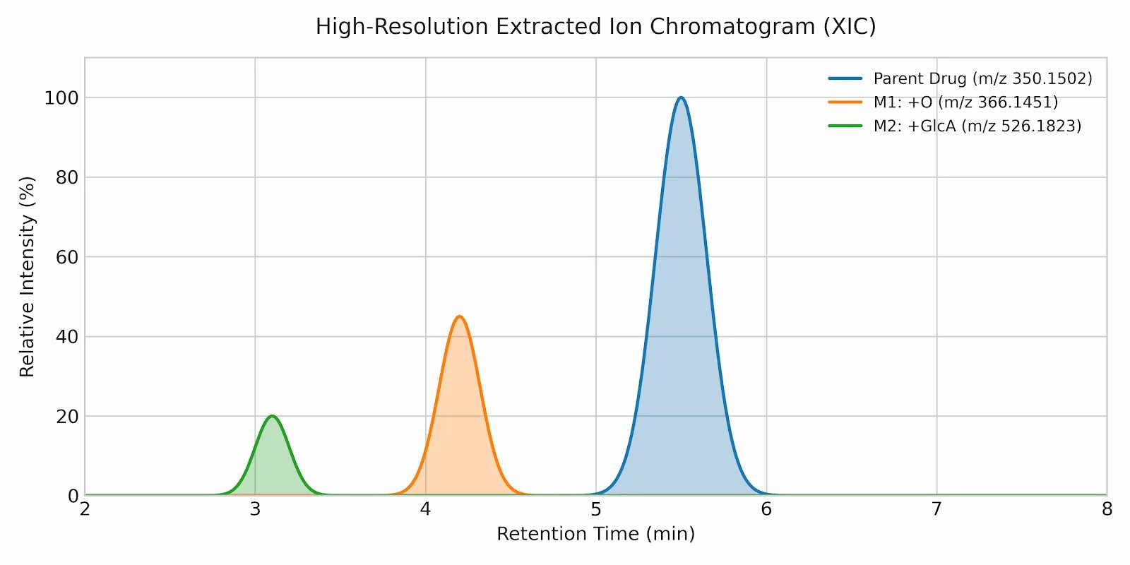
  • High-Resolution Extracted Ion Chromatograms (XIC): Clear visual overlays showing the exact chromatographic retention times of the parent drug alongside all newly identified metabolites, demonstrating their relative abundance and polarity shifts.
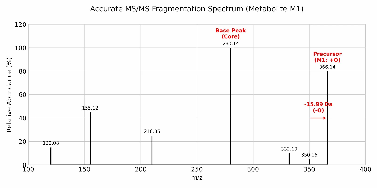
  • Accurate MS/MS Fragmentation Trees: Detailed structural mass maps showing the exact collision-induced fragment ions. We highlight the specific mass shifts (e.g., +15.9949 Da for oxidation) directly on the chemical backbone, proving exactly where the biotransformation occurred.
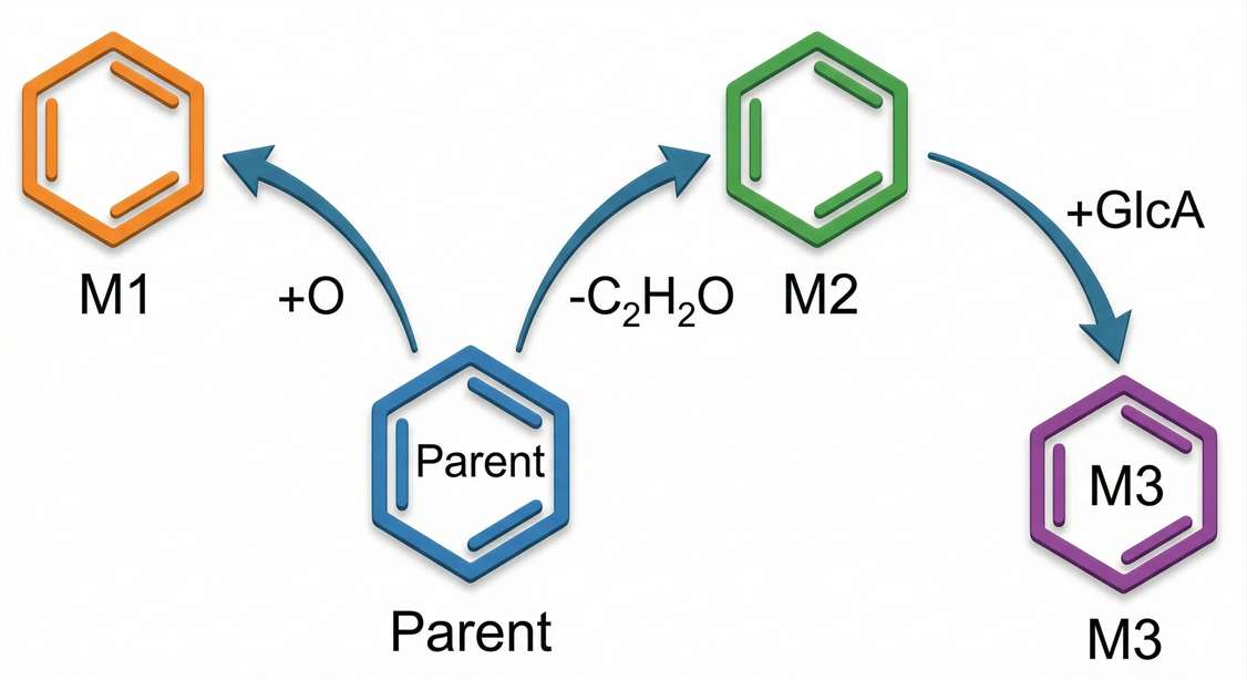
  • Proposed Metabolic Pathway Maps: A comprehensive, node-based flow chart illustrating the complete biotransformation network. This map uses directed edges to show how the parent compound systematically converts into its primary, secondary, and tertiary metabolic products over time.
High-Resolution Extracted Ion Chromatograms (XIC)
Accurate MS/MS Fragmentation Trees
Proposed Metabolic Pathway Maps

Standardized Sample Requirements for In Vitro Profiling

To ensure optimal in vitro incubation and maximum MS ionization efficiency, please adhere to the following submission guidelines. If your compound exhibits poor solubility or requires specific compound stability and degradation studies before incubation, please notify our technical team in advance.

  • Test Compound (Solid Powder): We recommend providing 1 to 5 mg shipped dry at room temperature. A purity strictly >95% is required. Please provide the exact molecular weight, salt form, and any known structural vulnerabilities to assist our initial mass spectrometry tuning.
  • Test Compound (Solution): Please provide at least 100 μL at a 10 mM stock concentration, shipped on dry ice (-20°C). It is absolutely critical to specify the exact solvent used (e.g., DMSO, Methanol). High DMSO concentrations can severely inhibit CYP enzymatic activity during microsomal incubation and must be accounted for.
  • Reference Standards (for Known Metabolites): If available, providing >1 mg of synthetic standards shipped on dry ice is highly recommended. Supplying synthetic standards for suspected downstream metabolites allows our team to perform definitive Level 1 structural confirmation and precise chromatographic alignment against your in vitro samples.

Case Study: Resolving Complex Metabolic Pathways

Source Paper

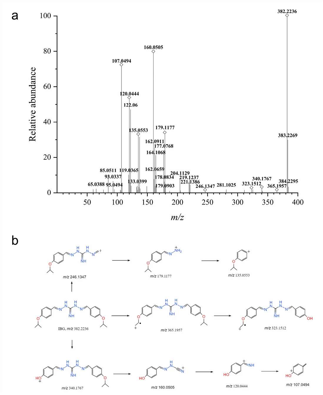
Metabolite Identification of Isopropoxy Benzene Guanidine in Rat Liver Microsomes by Using UHPLC-Q-TOF-MS/MS doi:10.3390/ijms24087313

Background

Identifying the precise biotransformation routes of a novel compound is an essential milestone for early de-risking. In this verified scientific scenario, researchers investigated Isopropoxy Benzene Guanidine (IBG), a novel derivative with promising antibacterial activity, to deeply understand its metabolic fate and clearance mechanisms in liver microsomes.

Methods

The parent compound (IBG) was incubated in vitro with rat liver microsomes supplemented with NADPH to trigger hepatic oxidative metabolism. Instead of standard targeted quantification, the quenched samples were analyzed using an untargeted UHPLC-Q-TOF-MS/MS platform. High-resolution product ion scanning was utilized to capture highly accurate fragmentation data across a wide mass range. Strict Mass Defect Filtering (MDF) algorithms were applied, establishing a narrow mass defect window centered around the parent molecule's core structure.

Results & Conclusion

The high-resolution HRMS profiling coupled with MDF successfully identified 7 distinct metabolites that would have been invisible to standard QqQ systems. By establishing the baseline fragmentation tree, scientists could confidently assign downstream mass shifts. By mapping these high-accuracy mass fragments against the parent structure, the research team successfully elucidated the primary metabolic pathways of IBG—revealing crucial routes including monohydroxylation, dihydroxylation, and specific O-dealkylation events.

This article is licensed under a Creative Commons Attribution 4.0 International License (CC BY 4.0).

Metabolite Identification of IBG via UHPLC-Q-TOF-MS/MS

Ready to Map Your Compound's Metabolic Fate?

Do not let unrecognized metabolic liabilities or hidden reactive intermediates derail your lead optimization phase. Share your compound's general class, target matrix, and suspected vulnerabilities with our bioanalytical experts. We will engineer a custom HR-LC-MS/MS profiling protocol to uncover its exact structural biotransformations and provide the definitive analytical data needed for successful molecular redesign.

REQUEST METABOLIC PROFILING DESIGN

Frequently Asked Questions

How does MetID differ from your standard single drug quantification service?

Standard quantification (single drug quantification) relies on Triple Quadrupole (QqQ) mass spectrometers operating in Multiple Reaction Monitoring (MRM) mode. This approach requires pre-existing knowledge of the exact molecule to measure its absolute concentration. In contrast, MetID uses High-Resolution Mass Spectrometry (HRMS), such as Q-TOF or Orbitrap platforms, to perform full-scan untargeted analysis. It searches for unknown molecules and focuses exclusively on qualitative structural elucidation rather than calculating absolute quantitative concentrations.

Can HR-LC-MS/MS effectively distinguish between positional isomers of a newly formed metabolite?

While HRMS provides the exact elemental formula with sub-ppm accuracy, it cannot differentiate positional isomers (e.g., hydroxylation occurring at the ortho, meta, or para position of a functional ring) purely by intact mass, as they have identical molecular weights. We resolve this complex challenge by optimizing the chromatographic stationary phase (using specialized columns like PFP or Phenyl-Hexyl) to physically separate the isomers based on subtle polarity differences. Once separated by retention time, we conduct an in-depth analysis of their distinct MS/MS fragmentation intensity ratios to assign the correct structural position.

What strategy do you employ if a compound clears too slowly in microsomes to generate detectable metabolite signals?

For exceptionally stable compounds with extremely low intrinsic clearance, a standard 60-minute microsomal incubation may yield insufficient metabolite mass for confident HRMS detection. In these challenging cases, we recommend transitioning to a hepatocyte relay assay, or utilizing specialized engineered recombinant CYP models that offer extended incubation viability. This allows us to artificially enrich the metabolite pool over several hours prior to sample extraction and high-resolution mass analysis.

Ready to Quantify Your Lead Compound or Metabolite?

Share your matrix type, sample count, and expected range—feasibility routing will confirm whether direct quantification is fit-for-purpose or method development is recommended.

inquiry
Online Inquiry