The Bioanalytical Revolution for Oligonucleotide Therapeutics
Oligonucleotide therapeutics — including antisense oligonucleotides (ASOs), small interfering RNAs (siRNAs), microRNAs (miRNAs), messenger RNAs (mRNAs), and aptamers — represent one of the fastest-growing and most versatile therapeutic modalities in modern biopharma. By directly targeting pre-mRNA, mRNA, or gene expression pathways, nucleic acid-based drugs enable the selective modulation of previously "undruggable" disease targets across oncology, rare genetic disorders, neurodegenerative conditions, and cardiovascular diseases. However, the successful clinical translation of therapeutic oligonucleotides hinges on robust, highly sensitive, and specific bioanalytical methods capable of accurately quantifying parent drugs and their active or inactive metabolites in complex biological matrices (plasma, serum, CSF, and tissue homogenates). At Creative Proteomics, our specialized DMPK bioanalysis platform offers comprehensive custom LC-MS/MS method development for novel chemical entities and complex nucleic acid drugs, supporting discovery PK, lead optimization, and IND-enabling safety studies.
Historically, bioanalysis of therapeutic oligonucleotides relied predominantly on ligand-binding assays (LBAs), such as hybridization ELISA, electrochemiluminescence (ECL), and stem-loop RT-qPCR. While hybridization LBAs offer excellent sensitivity (sub-ng/mL LLOQ), they suffer from major analytical limitations: (a) cross-reactivity with closely related truncated metabolites (e.g., 3'- or 5'-end 3'-N-1, N-2, or N-3 short-mer catabolites that differ by only a single nucleotide), (b) narrow dynamic range requiring extensive sample dilution, (c) requirement for custom-synthesized, high-affinity complementary hybridization probes for every new candidate, and (d) inability to characterize chemical modifications (e.g., phosphorothioate PS backbones, 2'-O-methyl 2'-OMe, 2'-fluoro 2'-F, 2'-O-methoxyethyl 2'-MOE, GalNAc conjugation, or lipid adducts). Liquid chromatography coupled with tandem mass spectrometry (LC-MS/MS) and high-resolution mass spectrometry (HRMS) has emerged as the gold standard platform for oligonucleotide bioanalysis, offering unambiguous mass-based specificity, wide linear dynamic range (3–4 orders of magnitude), and the unique capability to resolve intact drug candidates from structural impurities and shortened catabolites in a single assay run.
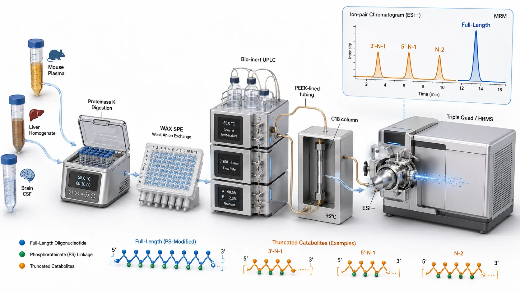 Figure 1: End-to-End LC-MS/MS Bioanalytical Architecture for Oligonucleotide Therapeutics — 3D Diagram
To overcome these limitations and mitigate translational risk, modern preclinical DMPK and IND-enabling studies require multi-component bioanalysis — the simultaneous, quantitative profiling of multiple molecular species (parent drugs, structural components, catabolites, and pharmacodynamic biomarkers) from a single biological sample aliquot. Operating across plasma and serum drug quantification as well as complex biological matrices analysis (including tumor tissue homogenates, CSF, bile, and cell lysates), multi-component workflows eliminate exposure misinterpretation, refine human PK predictions, and satisfy stringent regulatory expectations set forth by FDA, EMA, and ICH M10 bioanalytical guidelines.
Developing robust LC-MS/MS methods for oligonucleotide therapeutics requires overcoming severe physicochemical challenges inherent to nucleic acid chemistry. Oligonucleotides are highly polar, polyanionic macromolecules containing multiple negatively charged phosphate or phosphorothioate backbone groups. This polyanionic structure causes severe non-specific adsorption to metal surfaces in LC systems and glassware, complex charge-state distributions in electrospray ionization (ESI) negative mode, and intense adduct formation with sodium and potassium ions that dilutes analyte signal. Overcoming these hurdles requires specialized ion-pair reversed-phase chromatography (IP-RP-LC-MS), bio-inert LC hardware, and optimized sample extraction protocols utilizing sample preparation method development services tailored to biological matrices.
Physicochemical Properties and Key Bioanalytical Challenges of Nucleic Acids
Understanding the unique chemical architecture of synthetic oligonucleotides is prerequisite to establishing reliable bioanalytical workflows. Unlike hydrophobic small molecules or globular proteins, oligonucleotides possess extreme physical properties that govern their behavior during extraction, chromatographic separation, and mass spectrometric detection.
1. Polyanionic Backbone and Non-Specific Metal Adsorption
Synthetic ASOs and siRNAs typically range from 15 to 30 nucleotides in length (molecular weight ~5,000 to 10,000 Da) and carry 14 to 29 negative charges at physiological pH due to the phosphodiester or phosphorothioate (PS) backbone. This concentrated negative charge density drives strong electrostatic interactions with positively charged metal oxide sites on standard stainless steel LC components, analytical columns, injector needles, and glass autosampler vials. This non-specific metal adsorption causes severe peak tailing, carryover, poor peak area reproducibility, and total signal loss at sub-microgram concentrations. Mitigating non-specific binding requires: (a) bio-inert or PEEK-lined LC hardware and column hardware, (b) mobile phase additives such as chelating agents (e.g., EDTA, medronic acid, or citric acid) at micromolar levels, and (c) passivating LC flow paths prior to analytical runs.
2. Multiple Charge State Distribution and Alkali Metal Adduction in ESI
During negative-mode electrospray ionization (ESI-), polyanionic oligonucleotides undergo multiple deprotonation events, yielding a broad charge state distribution envelope (typically [M-5H]5- down to [M-15H]15- depending on length and modifier chemistry). Spreading the total ion current across 5–10 distinct charge state channels inherently reduces the signal intensity of any single precursor ion selected for MRM or PRM monitoring. Furthermore, endogenous sodium (Na+) and potassium (K+) ions present in biological extracts readily replace protons on the phosphate backbone, producing an abundance of alkali metal adducts ([M - nH + xNa](n-x)-) for each charge state. This adduct splitting severely fragments the analytical signal into multiple mass peaks, raising LLOQ levels. Effective bioanalysis demands volatile organic ion-pairing reagents in mobile phases to displace alkali counterions and collapse the charge state envelope into 1–2 dominant precursor ions.
3. Matrix Protein Binding and Complex Tissue Distribution
Therapeutic oligonucleotides exhibit high protein binding (>90–99% in plasma, predominantly to serum albumin and alpha-2-macroglobulin) driven by hydrophobic interactions with phosphorothioate backbones and 2'-modifications. In tissue homogenates (liver, kidney, spleen, brain/CSF), oligonucleotides bind tightly to cell nuclear matrix and intracellular proteins, making direct liquid-liquid extraction ineffective. Furthermore, oligonucleotide therapeutics heavily accumulate in target tissues (with liver and kidney tissue-to-plasma partition coefficients Kp > 10–50), whereas circulating plasma levels clear rapidly within hours post-dose. Accurately measuring tissue exposure and intracellular clearance requires specialized tissue homogenization, Proteinase K enzymatic digestion, and complex biological matrices analysis services capable of disrupting protein-nucleic acid complexes while preserving full-length drug integrity.
Ion-Pair Reversed-Phase (IP-RP-LC-MS) vs. Alternative Separation Modes
Chromatographic separation of highly polar, polyanionic oligonucleotides cannot be achieved using standard reversed-phase C18 methods without chemical modification of the flow stream. Ion-Pair Reversed-Phase Liquid Chromatography (IP-RP-LC-MS) is the premier separation technology utilized across discovery and regulated bioanalysis.
1. Mechanism of Ion-Pairing Reagents and Fluorinated Organic Modifiers
IP-RP-LC-MS utilizes volatile alkylamine ion-pairing (IP) agents — such as triethylamine (TEA), triethylammonium acetate (TEAA), dibutylamine (DBA), hexylamine (HA), or diisopropylethylamine (DIPEA) — added to the aqueous mobile phase A. The hydrophobic alkyl chain of the amine partitions into the stationary phase of a C18 or C8 column, while the positively charged ammonium head group faces the aqueous mobile phase. This forms a dynamic anion-exchange surface that electrostatically retains negatively charged oligonucleotides. Simultaneously, a volatile fluorinated alcohol organic modifier — most notably 1,1,1,3,3,3-hexafluoro-2-propanol (HFIP) or 1,1,1,3,3,3-hexafluoro-2-methyl-2-propanol (HFIP/HFTB) — is added to both mobile phases A and B (typically 10–100 mM HFIP combined with 5–15 mM TEA/DBA in water/methanol or water/acetonitrile).
HFIP serves three vital functions: (a) it acts as a weak acid buffer maintaining mobile phase pH around 8.0–8.5 (ensuring full deprotonation of HFIP and ion-pairing amines), (b) it dramatically enhances ESI negative-mode ionization efficiency through gas-phase charge transfer and droplet evaporation enhancement, and (c) it prevents alkali metal (Na+/K+) adduction by outcompeting inorganic cations during droplet formation. Optimizing the molar ratio of alkylamine to HFIP (e.g., 15 mM TEA / 100 mM HFIP or 10 mM DBA / 40 mM HFIP) is the single most critical parameter governing chromatographic resolution of full-length oligonucleotides from 3'- and 5'-truncated catabolites (e.g., N-1, N-2 species).
2. Column Chemistry, Temperature, and System Contamination Management
IP-RP separations require elevated column temperatures (50°C to 75°C) to denature self-complementary secondary structures (such as hairpin loops or double-stranded siRNA duplexes) into single strands, ensuring sharp, symmetrical chromatographic peaks. Stationary phases must feature wide-pore organosilica or hybrid particles (130Å to 300Å pore size, sub-2 µm or 2.5 µm particle size, e.g., C18, C8, or phenyl-hexyl) designed to withstand high pH (pH 8–10) and high temperatures without silica dissolution. A major operational challenge of IP-RP-LC-MS is persistent system contamination: ion-pairing alkylamines strongly adsorb onto LC tubing and MS ion source components, causing long-term suppression for positive-mode assays. Dedicated LC-MS instruments specifically allocated for negative-mode oligonucleotide bioanalysis are strongly recommended.
3. Alternative Modes: HILIC, Anion Exchange (AEX), and Non-IP Reversed-Phase
While IP-RP-LC-MS dominates the field, alternative separation modes offer distinct advantages in specific contexts:
- Hydrophilic Interaction Liquid Chromatography (HILIC): Retains polar oligonucleotides on zwitterionic or amide stationary phases using high organic mobile phases (70–85% ACN) without requiring alkylamine ion-pairing reagents. HILIC avoids system contamination and provides high MS sensitivity due to organic-rich spray conditions, but offers lower chromatographic resolution for single-nucleotide N-1 truncated catabolites compared to IP-RP.
- Anion-Exchange Chromatography (AEX): Separates oligonucleotides strictly based on charge state using strong anion exchange columns with salt gradients (e.g., NaCl or NaClO4). Excellent for preparative purification and purity profiling, but non-volatile salt mobile phases render AEX incompatible with direct online ESI-MS detection unless coupled to an online desalting module.
- Non-IP Reversed-Phase Chromatography: Employs specialized stationary phases (e.g., polar-embedded C18 or fluorinated phases) with high-organic/neutral pH mobile phases without ion-pairing reagents, avoiding mass spec contamination at the expense of resolution for long oligonucleotide chains.
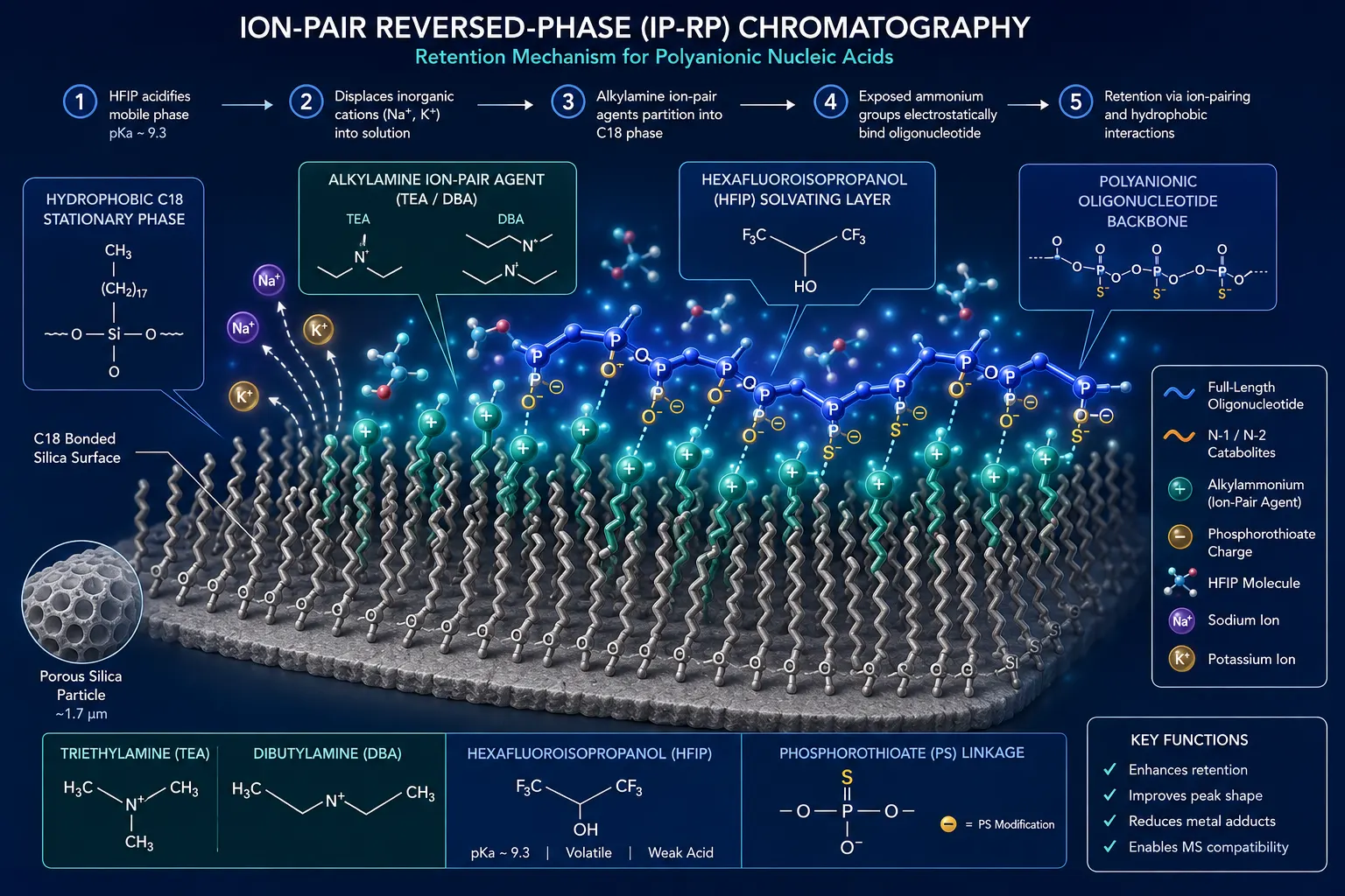 Figure 2: Ion-Pair Reversed-Phase (IP-RP) Dynamic Retention and Charge Neutralization Mechanism
Sample Preparation Strategies: SPE, Hybridization Capture, and Tissue Extraction
Biological matrices (plasma, serum, tissue homogenates) contain vast excesses of endogenous proteins, genomic DNA, RNA, and phospholipids that interfere with LC-MS quantification. Selecting a tailored sample extraction protocol is vital to achieve sub-ng/mL LLOQ limits while preserving analyte recovery.
1. Pretreatment: Disruption of Protein Binding via Proteinase K Digestion
Direct liquid-liquid extraction or protein precipitation (PPT) fails for oligonucleotide bioanalysis because polar nucleic acids co-precipitate with plasma proteins or remain bound to cellular tissue matrix, yielding recoveries below 10–20%. Effective pretreatment requires enzymatic protein digestion. Biological samples (10–100 µL plasma or tissue homogenates prepared in lysis buffer containing EDTA and TRIS) are incubated with Proteinase K (a broad-spectrum serine protease) at 50°C–55°C for 1 to 3 hours. Proteinase K completely digests matrix proteins (including albumin and nucleases) into small peptide fragments, releasing bound oligonucleotides into solution without degrading modified phosphorothioate backbones or 2'-modifications.
2. Solid-Phase Extraction (SPE): Weak Anion Exchange (WAX) and Reversed-Phase SPE
Following Proteinase K digestion, Solid-Phase Extraction (SPE) in 96-well plate format provides rapid, high-recovery sample cleanup. The most effective sorbent chemistries include:
- Weak Anion-Exchange (WAX) SPE: Utilizes polymeric sorbents functionalized with tertiary amine groups. At pH ~5–6, the sorbent is positively charged, selectively retaining polyanionic oligonucleotides via strong ionic interactions while neutral lipids, peptides, and small-molecule interferences are washed away with water and organic solvents. Elution is achieved at pH ~9–10 using volatile ammonium hydroxide/methanol solutions that deprotonate the sorbent, yielding recovery >85% and phospholipid removal >98%.
- Clarity OTX SPE: A specialized mixed-mode sorbent engineered specifically for synthetic oligonucleotides. Isolates ASOs, siRNAs, and catabolites from tissue homogenates with minimal lipid carryover and high LLOQ sensitivity.
3. Hybridization Assay Capture LC-MS/MS: Sub-ng/mL Sensitivity for Regulated Studies
For ultra-sensitive applications (e.g., low-dose CNS administration, CSF matrix, or pediatric micro-samples where LLOQ < 0.1–0.5 ng/mL is required), Hybridization Capture LC-MS/MS provides unprecedented selectivity. A biotinylated complementary DNA or LNA probe designed against the target oligonucleotide sequence is incubated with the biological sample. The probe hybridizes specifically with intact target drug molecules. The resulting duplex is captured on streptavidin-coated magnetic microbeads or 96-well plates. After rigorous washing to eliminate 99.9% of matrix interferences, the target oligonucleotide is thermally or chemically eluted and injected onto the IP-RP-LC-MS/MS system. Hybridization LC-MS combines the extreme sensitivity of hybridization capture with the unambiguous mass-resolving power of LC-MS/MS, resolving full-length drug from truncated N-1/N-2 catabolites.
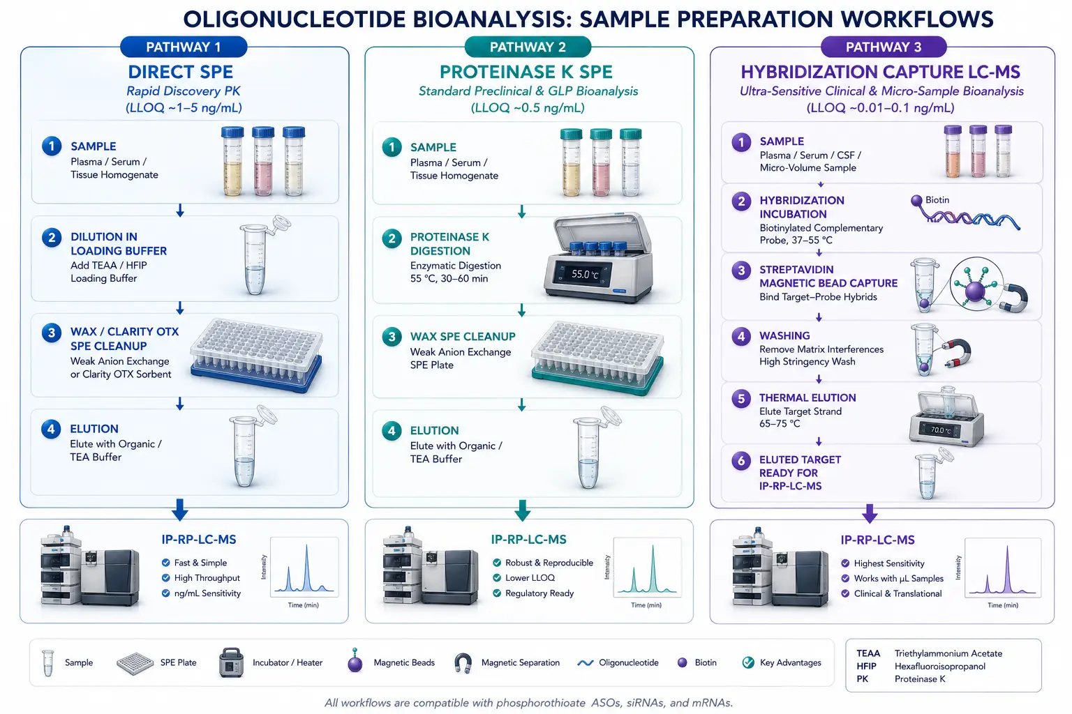 Figure 3: Multi-Stage Sample Extraction Workflows: Direct SPE vs. Proteinase K SPE vs. Hybridization Capture
Mass Spectrometry Detection: MRM, PRM, and Internal Standard Selection
Mass spectrometric detection provides the analytical specificity required to quantify target oligonucleotides amidst complex biotransformation products.
1. Triple Quadrupole MRM Transitions and Backbone Fragment Selection
Triple quadrupole mass spectrometers operated in negative ESI Multiple Reaction Monitoring (MRM) mode offer fast cycle times and wide dynamic range. Precursor ion selection targets the 2 or 3 most abundant charge states within the charge state envelope (e.g., [M-7H]7- or [M-8H]8- for a 20-mer ASO). Collision-Induced Dissociation (CID) fragmentation produces characteristic product ions:
- Phosphorothioate (PS) Backbone Fragments: CID of PS-modified oligonucleotides yields intense, highly reproducible universal product ions at m/z 95 (thiophosphate, [PO2S]-) and m/z 319 (phosphorothioate nucleotide fragments). Monitoring precursor charge states → m/z 95 provides ultra-sensitive MRM quantification for PS-modified ASOs.
- Sequence-Specific Oligonucleotide Fragments: CID of phosphodiester or mixed-backbone oligonucleotides yields sequence-specific w-type and a-B-type fragment ions. Selecting sequence-specific product ions provides maximum specificity against endogenous RNA/DNA fragments.
2. High-Resolution Mass Spectrometry (HRMS): PRM and Full-Scan MS1 Profiling
High-Resolution Mass Spectrometry (HRMS) on Orbitrap or Q-TOF platforms operating at resolving power > 60,000 to 120,000 offers major advantages for oligonucleotide bioanalysis. Operating in Parallel Reaction Monitoring (PRM) or full-scan MS1 mode, HRMS allows post-acquisition extraction of target mass chromatograms with sub-5 ppm mass accuracy. HRMS resolves isotopic envelopes of intact full-length oligonucleotides from single-dalton truncated metabolites (e.g., resolution of 3'-N-1, 5'-N-1, deamination products, and oxidation products) without requiring pre-selected MRM fragmentation transitions, making HRMS the gold standard for comprehensive metabolite identification (MetID) and catabolite profiling.
3. Internal Standard (IS) Strategy: SIL-IS vs. Elongated Analog IS
The choice of internal standard (IS) is critical to correct for extraction variability, non-specific metal adsorption, and IP-RP ESI ion suppression. Three primary IS strategies are deployed:
- Stable Isotope-Labeled IS (SIL-IS): Synthesized by incorporating 13C- and 15N-labeled nucleotides into the target sequence. SIL-IS is chemically identical to the target analyte, co-elutes perfectly, and experiences identical matrix effects and adsorption losses, providing CV < 5% precision. SIL-IS is the gold standard for regulated GLP studies.
- Elongated / Truncated Analog IS: A synthetic oligonucleotide with identical sequence and backbone chemical modifications, elongated or shortened by 1 to 2 nucleotides (e.g., N+1 or N+2 sequence). Exhibits near-identical extraction recovery and chromatographic behavior while being fully resolved in mass domain.
- Fully Modified Non-Target Analog IS: A non-homologous oligonucleotide carrying matching 2'-modifications and PS backbone chemistry. Available at lower cost for early discovery PK screening.
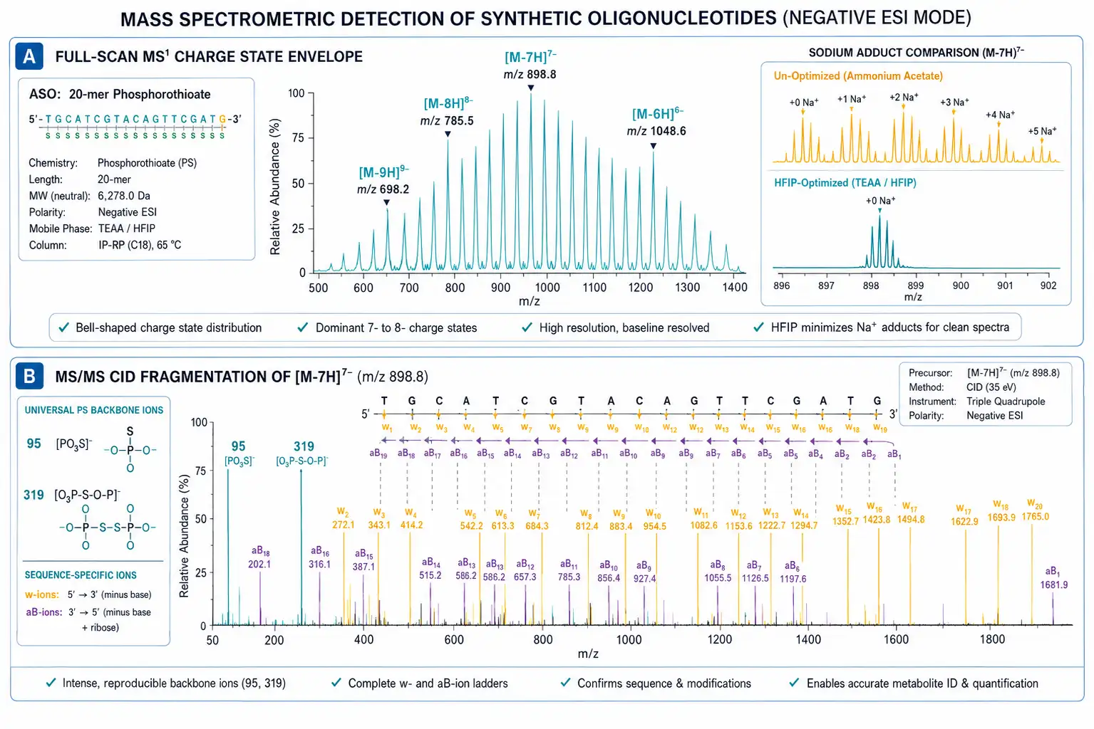 Figure 4: Negative-Mode ESI Charge State Envelope and MS/MS CID Fragmentation Spectrum
Regulatory Compliance & Method Validation Under ICH M10 Guidelines
Bioanalytical methods supporting IND-enabling safety studies, GLP toxicology, and clinical trials must be rigorously validated per the ICH M10 global guideline. Validating oligonucleotide LC-MS assays requires specific adaptations to account for nucleic acid chemistry.
1. Selectivity and Catabolite Specificity (N-1, N-2 Resolution)
Selectivity must be evaluated in at least six individual matrix lots (including hemolyzed and lipemic matrix lots). Blank matrix must demonstrate no significant interference (<20% of LLOQ response and <5% of IS response) at the target retention time. Crucially, method specificity must demonstrate baseline chromatographic or mass-based resolution from short-mer catabolites (3'-N-1, 5'-N-1, N-2 species). Spiked QC samples containing N-1 catabolites at 10–50% of parent drug concentration must demonstrate that the catabolite does not contribute >5% bias to the parent quantification.
2. Linearity, Accuracy, Precision, and Weighting Factors
Calibration curves must contain at least 6 to 8 non-zero calibrator levels spanning the intended analytical range (e.g., 0.5 ng/mL to 1,000 ng/mL in plasma or 5 ng/g to 10,000 ng/g in liver tissue). Weighted linear regression (1/x² weighting) is standard to correct for heteroscedasticity across the calibration range. Within-run and between-run accuracy must be within 85–115% of nominal concentration (80–120% at LLOQ), with precision (CV) ≤ 15% (≤ 20% at LLOQ) across Low, Mid, and High QC levels evaluated in at least 3 independent validation runs.
3. Matrix Factor, Carryover, and Passivation Validation
Matrix effects evaluated across 6 individual lots must demonstrate IS-normalized matrix factor CV ≤ 15%. Because ion-pairing reagents can interact with matrix components, matrix factor evaluation is essential. Carryover must be rigorously evaluated by injecting blank matrix samples immediately after the highest calibrator (ULOQ); carryover signal must not exceed 20% of LLOQ. Passivation procedures (e.g., injecting high-concentration sacrificial oligonucleotide or medronic acid solution prior to analytical runs) must be documented in formal SOPs.
4. Stability Evaluation Across Processing and Storage
Stability must be fully established under all handling conditions:
- Bench-Top Stability: Plasma/tissue homogenate stability at room temperature and 4°C for ≥ 4–24 hours (verifying no enzymatic degradation by residual nucleases).
- Freeze-Thaw Stability: At least 3 to 5 freeze-thaw cycles (-80°C to room temperature).
- Long-Term Storage Stability: Matrix storage stability at -80°C for at least 6 to 12 months.
- Autosampler Stability: Processed extract stability in IP-RP mobile phase in autosampler (4°C) for ≥ 48–72 hours (confirming no non-specific vial adsorption or HFIP/TEA evaporation). Supported by our full and partial method validation services under ISO/IEC 17025 accreditation, sponsors receive complete, audit-ready validation packages.
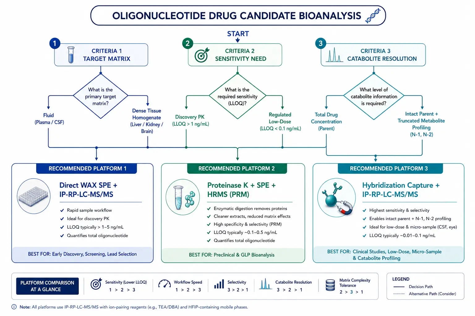 Figure 5: Decision Tree for Selecting Bioanalytical Methodologies for Oligonucleotide Therapeutics
Decision Matrix: Hybridization LBA vs. IP-RP-LC-MS/MS vs. Hybrid Capture LC-MS/MS
Selecting the optimal bioanalytical strategy for oligonucleotide drug candidates depends on development stage, sensitivity requirements, and catabolite profiling needs. The following decision matrix compares the primary bioanalytical platforms across key technical parameters:
| Parameter |
Hybridization LBA (ELISA/ECL) |
Direct IP-RP-LC-MS/MS |
Hybrid Capture LC-MS/MS |
| Primary Analytical Scope |
Total oligonucleotide concentration in fluid matrices |
Simultaneous parent & truncated catabolite profiling |
Ultra-sensitive parent & catabolite quantification |
| Analytical Specificity |
Moderate; cross-reacts with N-1/N-2 truncated metabolites |
High; mass-based resolution of single-nucleotide catabolites |
Ultra-High; sequence hybridization + mass specificity |
| Sensitivity (Typical LLOQ) |
High (0.01 – 0.1 ng/mL) |
Moderate to High (0.5 – 5 ng/mL) |
Ultra-High (0.01 – 0.1 ng/mL) |
| Dynamic Range |
Narrow (1 – 2 orders of magnitude) |
Wide (3 – 4 orders of magnitude) |
Wide (3 – 4 orders of magnitude) |
| Reagent Requirements |
Custom complementary probe & antibody reagents required |
Off-the-shelf IP reagents (HFIP/TEA); no custom probes |
Custom biotinylated hybridization probe required |
| Tissue Matrix Performance |
Vulnerable to tissue matrix binding & background interferences |
High performance with Proteinase K + SPE cleanup |
Exceptional cleanup; captures target from tissue homogenates |
| Development Timeline |
4 – 8 weeks (probe synthesis & assay optimization) |
1 – 2 weeks (rapid LC-MS method tuning) |
2 – 4 weeks (probe synthesis & LC-MS optimization) |
| Regulatory Status (ICH M10) |
Widely accepted for clinical PK |
Gold standard for preclinical DMPK, MetID, and GLP tox |
Preferred for clinical low-dose PK & micro-sample bioanalysis |
Implementing IP-RP-LC-MS/MS or Hybrid Capture LC-MS/MS early in preclinical drug development ensures that PK/PD modeling is grounded in true parent drug exposure rather than confounded catabolite signals. Supported by Creative Proteomics' expert DMPK bioanalysis team, sponsors receive custom, regulatory-compliant analytical methods tailored to the exact chemistry of their RNA and ASO drug candidates.
Frequently Asked Questions
Why is ion-pairing (IP) required for LC-MS analysis of synthetic oligonucleotides?
Synthetic oligonucleotides possess a polyanionic phosphate or phosphorothioate backbone carrying 15–30 negative charges, making them extremely polar and unretained on standard C18 columns. Volatile ion-pairing alkylamines (e.g., TEA, DBA) added to mobile phases dynamically bind to the C18 stationary phase and electrostatically retain the negatively charged nucleic acids, enabling high-resolution chromatographic separation.
How do fluorinated modifiers like HFIP improve negative ESI-MS sensitivity for oligonucleotides?
Hexafluoroisopropanol (HFIP) acts as a volatile weak acid buffer (pH ~8.0) that enhances electrospray droplet surface evaporation and gas-phase charge transfer. Crucially, HFIP outcompetes inorganic cations (Na+ and K+), preventing alkali metal adduction and collapsing the broad charge state distribution into 1–2 dominant precursor ion channels, directly increasing signal intensity and LLOQ sensitivity.
How can non-specific metal adsorption be prevented during oligonucleotide LC-MS runs?
Oligonucleotide phosphate backbones bind strongly to metal oxide sites on stainless steel LC tubing, columns, and needles. Prevention strategies include: (a) utilizing bio-inert or PEEK-lined LC hardware, (b) adding micromolar chelating agents (e.g., medronic acid or EDTA) to mobile phases, and (c) passivating LC flow paths with sacrificial oligonucleotide injections prior to analytical runs.
Why is Proteinase K digestion necessary for tissue homogenate sample extraction?
Oligonucleotides bind tightly (>95–99%) to plasma and cellular matrix proteins. Direct solvent precipitation or liquid extraction fails because nucleic acids co-precipitate with proteins. Incubating tissue homogenates or plasma with Proteinase K at 50°C–55°C digests matrix proteins into small peptides, completely releasing bound target oligonucleotides for subsequent SPE extraction.
What is the difference between direct SPE and Hybridization Capture LC-MS/MS?
Direct SPE (e.g., WAX or Clarity OTX SPE) uses anion-exchange sorbents for rapid, high-throughput cleanup, achieving LLOQs around 0.5–5 ng/mL. Hybridization Capture LC-MS/MS uses biotinylated complementary DNA/LNA probes on magnetic beads to selectively capture target oligonucleotides from complex matrices, achieving ultra-high sensitivity (LLOQ 0.01–0.1 ng/mL) for low-dose micro-samples.
How are truncated catabolites (3'-N-1, 5'-N-1) resolved from full-length oligonucleotides?
Truncated catabolites differ by a single nucleotide (~300 Da mass difference). Baseline chromatographic separation is achieved using optimized IP-RP gradients (e.g., DBA/HFIP or TEA/HFIP mobile phases on high-temperature C18/C8 columns at 60°C–65°C), combined with High-Resolution Mass Spectrometry (Orbitrap/Q-TOF PRM) or high-mass-accuracy MRM monitoring.
What internal standards are recommended for regulated oligonucleotide bioanalysis?
Stable Isotope-Labeled Internal Standards (SIL-IS, containing 13C and 15N isotopes) are the gold standard for regulated GLP studies, as they co-elute perfectly and experience identical matrix effects. When SIL-IS is unavailable, elongated or shortened sequence analogs (e.g., N+1 or N+2 oligonucleotides with matching backbone chemical modifications) are highly effective surrogates.
Do oligonucleotide LC-MS methods comply with ICH M10 regulatory guidelines?
Yes. Bioanalytical LC-MS methods for oligonucleotides supporting GLP safety studies undergo full ICH M10 validation, independently establishing selectivity, linearity (1/x² weighting), within-run and between-run accuracy (85–115%), precision (CV ≤15%), matrix factor consistency (CV ≤15% across 6 lots), carryover, and long-term storage stability.
References
Related Services


