ISO 17025–ACCREDITED LABORATORY ENVIRONMENT

GSH Trapping Assay Services for Reactive Metabolite Screening

Finding toxicity risks late in the drug development lifecycle is a devastating and costly failure for any biotech pipeline. Our high-resolution GSH trapping assay provides a critical safety de-risking step for your early discovery efforts, allowing you to identify, map, and mitigate covalent binding liabilities long before candidate nomination.

We leverage state-of-the-art high-resolution liquid chromatography-tandem mass spectrometry (LC-HRMS/MS) paired with advanced bioinformatics. This combination cuts through the immense biological noise of in vitro incubations to confidently isolate trace-level reactive intermediates.

Predictive Risk Management

Identify hidden bioactivation pathways early to prevent drug-induced liver injury (DILI) liabilities.

Advanced Data Mining

Utilize Mass Defect Filtering (MDF) to accurately isolate low-abundance adducts from complex matrix backgrounds.

Bespoke Method Development

Customized incubation and quenching protocols designed specifically for challenging or novel chemical entities.

Biochemical Challenge Science of GSH Trapping Standardized Workflow Bioinformatics (MDF) Demo Results Sample Requirements Why Choose Us Case Study Related ServicesFAQ

The Biochemical Challenge: Why Screen for Reactive Metabolites?

During the standard phase I metabolism in the liver, cytochrome P450 (CYP) enzymes attempt to make lipophilic drugs more water-soluble for excretion. However, this process frequently creates unintended, highly unstable chemical byproducts known as reactive metabolites.

Because these intermediates are highly electrophilic (electron-deficient), they act like biological magnets. They aggressively seek out electron-rich targets within the cell, leading to irreversible covalent binding with essential cellular macromolecules such as proteins, DNA, and lipids.

This covalent modification can trigger severe cellular stress, initiate immune-mediated hypersensitivity reactions, or cause direct cellular necrosis. Historically, idiosyncratic drug-induced liver injury (DILI) caused by these reactive metabolites has been a leading reason for the withdrawal of approved drugs from the market and the termination of promising clinical trials.

Detecting these fleeting intermediates directly is virtually impossible due to their extremely short half-lives. Therefore, strategic in vitro screening using trapping agents is absolutely mandatory for proactive safety profiling.

The Science of Glutathione (GSH) Trapping: Hard vs. Soft Electrophiles

Not all reactive metabolites are created equal, which is why selecting the correct trapping agent is essential. In chemical terms, reactive intermediates are classified by Pearson's Hard and Soft Acids and Bases (HSAB) theory.

Most drug bioactivation pathways—such as the formation of epoxides, quinones, quinone imines, and Michael acceptors—produce what are known as "soft" electrophiles. Glutathione (GSH) is a naturally occurring tripeptide that contains a highly reactive thiol (-SH) group. This thiol group acts as a powerful "soft" nucleophile.

When we introduce an excess of GSH into an in vitro liver microsome incubation, it acts as a molecular sponge. It safely and rapidly captures these dangerous soft electrophiles before they can bind to microsomal proteins.

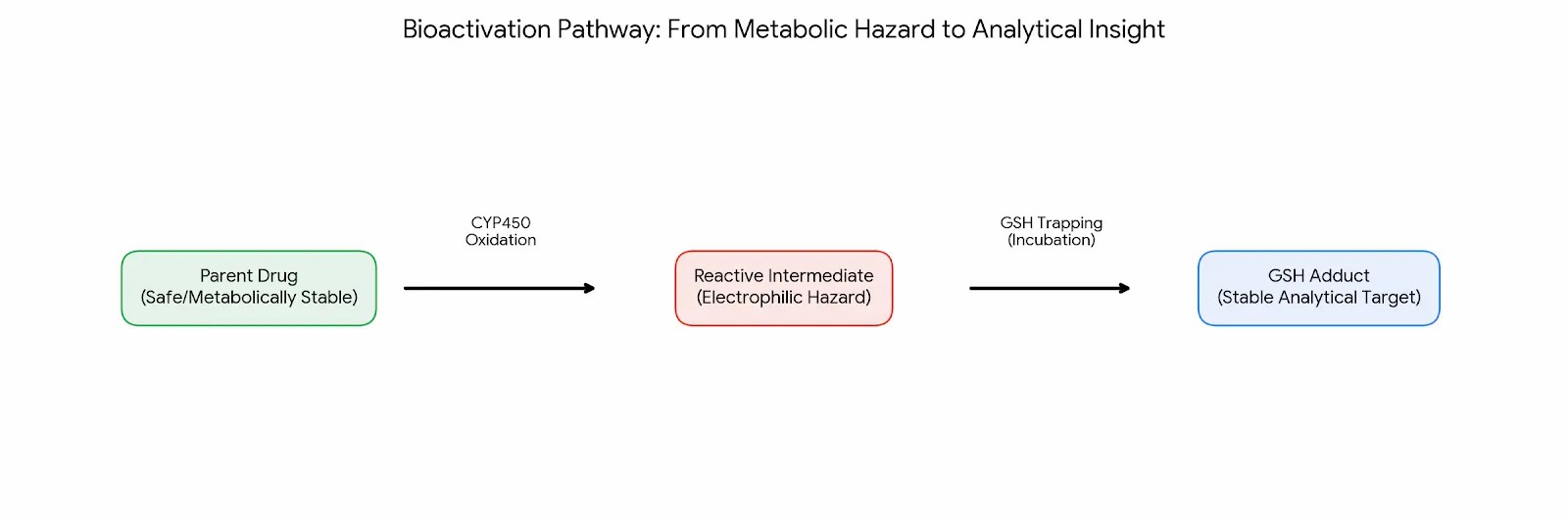
The resulting GSH-conjugate (adduct) is a highly stable compound. By stabilizing the threat, we transform an undetectable, fast-acting hazard into a stable analytical target that can be easily quantified and structurally characterized using our mass spectrometry platforms.

If your candidate is suspected of forming "hard" electrophiles (like certain aldehydes), our platform can seamlessly adapt to alternative trapping agents such as potassium cyanide (KCN) or methoxylamine.

Our Standardized Yet Adaptable GSH Trapping Workflow

Capturing unstable metabolites requires precision timing and carefully tuned incubation conditions. Our workflow is engineered to maximize sensitivity while maintaining enzyme viability.

Pre-Incubation Matrix Optimization

We rigorously evaluate your test compound's solubility. High concentrations of organic solvents like DMSO can rapidly inhibit CYP enzyme activity. We optimize the solvent ratios to ensure the parent drug remains in solution without suppressing the metabolic reaction.

In Vitro Bioactivation

As a core assay of our in vitro drug metabolism profiling, the compound is incubated at physiological temperatures with pooled human liver microsomes (HLM) or S9 fractions, supported by a continuous NADPH regenerating system, and an overwhelming molar excess of reduced GSH.

Precise Reaction Quenching

Reactions are terminated at highly specific time points using cold organic solvents, precipitating the proteins and locking the stable GSH adducts in their current state.

High-Resolution LC-MS/MS Data Acquisition

The processed samples are injected into high-resolution Orbitrap or Time-of-Flight (TOF) mass spectrometers. Data is collected using Data-Dependent Acquisition (DDA) to ensure both accurate precursor mass measurement and deep fragmentation data for structural mapping.

Advanced Bioinformatics: Mass Defect Filtering (MDF) to Eliminate Matrix Noise

The greatest challenge in reactive metabolite screening is not simply running the mass spectrometer—it is finding the data. A standard liver microsome incubation contains thousands of endogenous lipids, peptides, and buffer components. In this massive sea of biological noise, the target GSH adduct might represent less than 1% of the total ion current. Searching for it manually is unreliable and prone to false negatives.

This is where our bioinformatics expertise provides a definitive advantage. We utilize automated Mass Defect Filtering (MDF) to dramatically clean the data.

Every element has a precise fractional mass (e.g., Hydrogen is not exactly 1.0; it is 1.0078). Because drugs and their GSH adducts contain specific ratios of carbon, hydrogen, nitrogen, and sulfur, they exhibit a unique "mass defect" compared to the generic biological background.

When a parent drug binds to GSH, its mass increases by exactly 307.0838 Da (the mass of the added GSH moiety minus the displaced atoms). Our MDF algorithms mathematically establish a tight mass defect window around the parent drug. The software then scans the entire chromatogram and subtracts any signal that does not perfectly match the expected mass defect shift of a GSH conjugation.

This digital filtration strips away up to 95% of the endogenous matrix noise. The result is a brilliantly clean chromatogram where only the true, drug-related reactive adducts remain visible, allowing our scientists to detect even the most elusive, low-abundance toxic intermediates.

Comprehensive Demo Results: Translating Data into Structural Insight

We understand that early discovery teams do not want a raw list of molecular weights; they need actionable structural intelligence to guide medicinal chemistry. Our reporting format is built to provide maximum clarity.

  • Extracted Ion Chromatograms (XIC): Clean, noise-free visual graphs that clearly separate the remaining parent drug from the newly formed reactive adducts over the chromatography timeline.
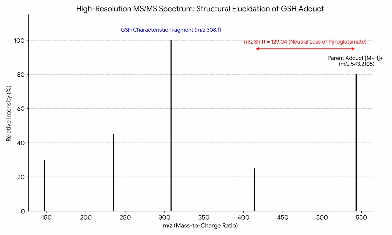
  • Neutral Loss Scanning Verification: Confirmation of the GSH adduct by identifying the characteristic loss of 129 Da (cleavage of the pyroglutamic acid moiety) in the fragmentation spectra.
  • High-Resolution MS/MS Structural Elucidation: Detailed fragmentation maps that pinpoint the exact location on the parent molecule where the GSH molecule attached.
  • Bioactivation Pathway Maps: Easy-to-understand visual diagrams illustrating the step-by-step metabolic transformation from the safe parent structure to the electrophilic hazard.
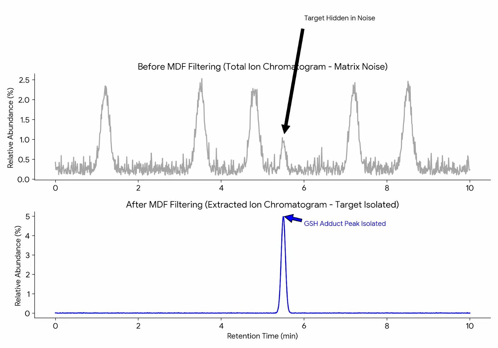
Extracted Ion Chromatograms of GSH adducts
High-Resolution MS/MS Structural Elucidation Spectra
Bioactivation Pathway Diagram

Sample Submission Requirements for High-Throughput Screening

To ensure the integrity of the assay and the accuracy of the mass spectrometry data, please adhere to our strict sample handling parameters.

Sample Type Minimum Recommended Input Concentration Shipping Condition
Test Compound (Powder) Minimum 1-5 mg required. N/A Ship at room temperature or on dry ice, protected from light.
Test Compound (Solution) Minimum 100-200 µL volume. Must be prepared at ≥ 10 mM concentration in high-purity DMSO. Ship immediately on dry ice.

Handling Note: Please provide any known chemical stability issues or light sensitivity warnings prior to shipping so our laboratory staff can prepare the appropriate amber vials and temperature-controlled reception protocols.

Why Choose the Creative Proteomics DMPK Platform?

Finding the right outsourcing partner for early de-risking requires looking beyond standard analytical testing capabilities.

Service Dimension Creative Proteomics DMPK Platform Conventional Analytical Testing
Structural Insight vs. Raw Data Output Our platform is staffed by structural elucidation experts who provide detailed interpretations of the fragmentation pathways through advanced metabolite identification (MetID) techniques, telling you exactly where the molecule is breaking down. Conventional analytical labs often deliver basic mass-shift reports.
De Novo Method Architecture Through our custom LC-MS/MS method development, we specialize in building bespoke extraction and chromatography methods from scratch to suit your unique chemical entity. Rigid, one-size-fits-all testing pipelines fail when confronted with highly lipophilic or complex novel structures.
Dual-Track Scalability We possess the agility to perform an intensive, multi-week mechanistic deep-dive on a single problematic lead compound, while also maintaining the high-throughput infrastructure necessary to screen a 500-compound library over a single weekend. Often restricted by fixed batch sizes and limited flexibility.

Research Case Study: Assessing Bioactivation Risks in Target Tissues via LC-HRMS

Background

Conventional in vitro screening sometimes fails to fully simulate metabolic activation within complex biological tissues. This study was conducted to validate a novel analytical strategy for the precise identification of reactive metabolites formed in situ within target tissues, providing a higher-fidelity assessment of exposure risks.

Methods

Researchers analyzed samples containing Glutathione (GSH) trapping agents using the Thermo Scientific Q Exactive high-resolution mass spectrometer. Advanced data mining was performed using Xcalibur and Compound Discoverer software, with a primary focus on leveraging Mass Defect Filtering (MDF) and Neutral Loss Scanning to cut through endogenous interferences.

Results

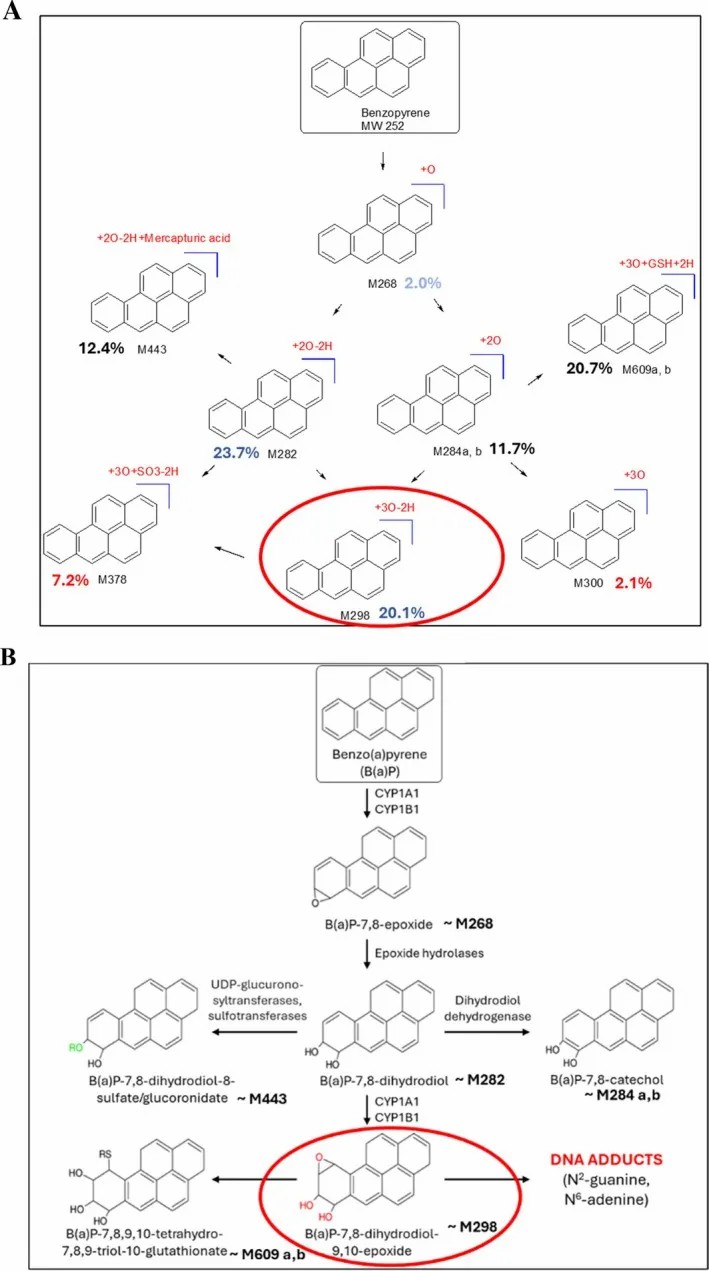
As documented in Figure 4 of the referenced literature, the study presented a comprehensive analytical workflow for reactive metabolite identification. By comparing the precise molecular weight shifts (accurate to four decimal places) between parent compounds and their respective GSH adducts, researchers successfully isolated and locked adduct signals generated through oxidative bioactivation.

Conclusion

This high-resolution mass spectrometry-based workflow effectively detects extremely low-abundance reactive intermediates. Furthermore, it characterizes specific covalent binding sites via MS2 characteristic fragments, providing a rigorous scientific basis for the structural de-risking and optimization of early-stage drug molecules.

Source Verification: Adapted from a 2025 Open Access study on target tissue metabolism and bioactivation assessment. View the Original CC-BY 4.0 Research Reference

Bioactivation identification workflow using HRMS and MDF (Reference: Figure 4)

Frequently Asked Questions (FAQ)

1. Why do we primarily use GSH instead of other trapping agents?

GSH is biologically relevant because it is the most abundant intracellular antioxidant in the human liver. Chemically, its thiol group makes it an excellent trap for "soft" electrophiles (like epoxides and quinones), which account for the vast majority of reactive metabolites in drug discovery.

2. Can this assay detect "hard" electrophiles?

No, GSH is poor at capturing hard electrophiles (like certain reactive aldehydes). If your compound’s structure suggests the formation of hard electrophiles, we will pivot our protocol to use hard nucleophile trapping agents such as potassium cyanide (KCN) or methoxylamine.

3. Does the formation of a GSH adduct guarantee clinical toxicity?

Absolutely not. The in vitro GSH trapping assay is a highly sensitive, exaggerated system designed to highlight potential risk. Many safe, marketed drugs form trace amounts of reactive metabolites. The assay is used for comparative ranking among your lead candidates to select the one with the lowest overall covalent binding burden.

4. How does your mass defect filtering (MDF) improve turnaround times?

Without MDF, a senior scientist might spend days manually combing through complex chromatograms looking for tiny mass shifts, which delays your project. MDF automates this digital subtraction, highlighting only the relevant adduct peaks in minutes. This allows our experts to spend their time on structural elucidation rather than data hunting, significantly accelerating your final report delivery.

5. At what stage of drug discovery should we utilize this screening?

We strongly recommend integrating GSH trapping assays during the Lead Optimization phase. Identifying a covalent binding liability early allows chemists to rationally redesign the molecule. Waiting until candidate selection or preclinical safety toxicology studies can result in massive financial losses if hepatotoxicity forces project termination.

Ready to Quantify Your Lead Compound or Metabolite?

Share your matrix type, sample count, and expected range—feasibility routing will confirm whether direct quantification is fit-for-purpose or method development is recommended.

inquiry
Online Inquiry