The Paradigm Shift: Why Single-Concentration Metrics Fail in Novel Modality DMPK
In classical small-molecule drug development, pharmacokinetic (PK) evaluation relies on a straightforward,
well-established paradigm: quantifying a single parent drug concentration in plasma or serum over time. That single
concentration metric directly informs systemic clearance, volume of distribution, terminal elimination half-life, and
exposure-driven safety margins. However, over the past decade, the biopharmaceutical pipeline has undergone a
fundamental structural transformation. Emerging modalities — including Antibody-Drug Conjugates (ADCs), Targeted
Protein Degraders (PROTACs/TPDs and Molecular Glues), bispecific/multispecific antibodies, and lipid nanoparticle
(LNP)-encapsulated RNA/oligonucleotide therapeutics — no longer fit this single-analyte bioanalytical framework. At
Creative Proteomics, our advanced DMPK platform provides specialized custom multi-analyte drug panel development and
comprehensive bioanalytical solutions engineered specifically to address the structural and pharmacokinetic
multi-dimensionality of novel drug candidates.
Emerging modalities are inherently multi-component, heterogeneous systems. An ADC, for instance, is not a homogenous
single molecule in vivo; upon intravenous administration, it rapidly transforms into a dynamic mixture of intact
conjugated antibody species with varying Drug-to-Antibody Ratios (DAR), total antibody, total payload, and catabolized
free payload. Similarly, a PROTAC bifunctional molecule undergoes complex ternary complex assembly with a target
protein and an E3 ubiquitin ligase, while simultaneously generating dealkylated, hydrolyzed, or oxidative
biotransformation products that can act as competitive antagonists or toxic off-target byproducts. Evaluating such
complex molecules using a single "total drug" or "total antibody" measurement creates profound analytical blind spots.
A stable total antibody concentration profile can easily mask rapid payload deconjugation in circulation, leading to
severe off-target systemic toxicity (such as neutropenia or thrombocytopenia) despite apparent therapeutic drug
levels. Conversely, measuring only total payload fails to distinguish active antibody-bound payload targeted to tumor
tissue from free payload circulating uncontrollably in systemic circulation.
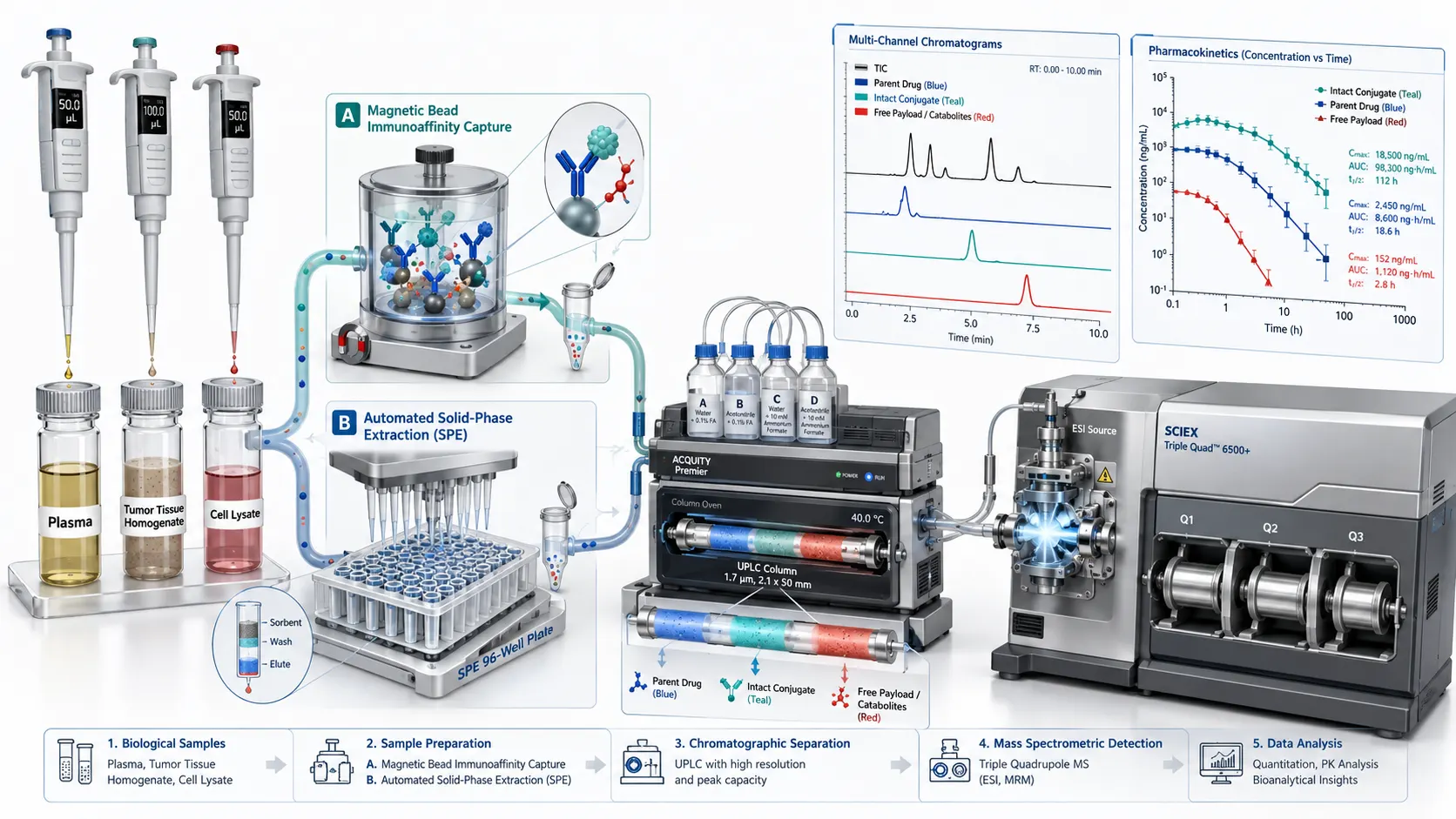 Figure 1: Multi-Component Bioanalytical Architecture for Emerging Therapeutic Modalities — 3D Diagram
To overcome these limitations and mitigate translational risk, modern preclinical DMPK and IND-enabling studies
require multi-component bioanalysis — the simultaneous, quantitative profiling of multiple molecular species (parent
drugs, structural components, catabolites, and pharmacodynamic biomarkers) from a single biological sample aliquot.
Operating across plasma and serum drug
quantification as well as complex biological matrices
analysis (including tumor tissue homogenates, CSF, bile, and cell lysates), multi-component workflows eliminate
exposure misinterpretation, refine human PK predictions, and satisfy stringent regulatory expectations set forth by
FDA, EMA, and ICH M10 bioanalytical guidelines.
Modality-Specific Bioanalytical Challenges and Multi-Component Requirements
Each novel therapeutic drug class introduces distinct biochemical structural features, biotransformation pathways,
and disposition mechanisms that dictate specific bioanalytical profiling strategies. Understanding these
modality-specific disposition profiles is prerequisite to designing robust, regulatory-compliant multi-component assay
panels.
1. Antibody-Drug Conjugates (ADCs): Resolving DAR Distribution, Payload Deconjugation, and Bystander Effect
ADCs combine the exquisite target specificity of monoclonal antibodies with the cytotoxic potency of small-molecule
payloads (such as auristatins [MMAE/MMAF], maytansinoids [DM1/DM4], or topoisomerase I inhibitors [DXd/SN-38]) via
cleavable or non-cleavable linkers. Upon intravenous administration, ADCs undergo systemic distribution alongside
continuous chemical and enzymatic cleavage in plasma and tissues. A complete ADC pharmacokinetic profile demands the
simultaneous tracking of at least three core analytes: conjugated antibody (antibody carrying at least one payload
molecule), total antibody (conjugated plus fully deconjugated naked antibody), and free payload (including active
catabolites such as Cys-linker-payload or Lys-linker-payload adducts).
Quantifying only total antibody provides zero insight into linker stability or payload clearance. If a linker is
labile, the free payload concentration spikes in systemic circulation, driving dose-limiting toxicities (e.g.,
neutropenia, interstitial lung disease, or ocular toxicity) while total antibody concentrations remain deceptively
stable. Furthermore, characterization of DAR distribution dynamics (from DAR 8 down to DAR 0 over time) using
high-resolution LC-MS/MS or hybrid LBA-LC-MS/MS workflows is essential to understand whether clearance is driven by
hepatic receptor-mediated endocytosis, FcRn recycling, or premature payload shedding. For ADCs utilizing
membrane-permeable payloads designed for bystander killing, multi-component analysis of intracellular vs.
extracellular payload in tumor tissue is critical. Our specialized ADCs DMPK and bioanalytical services integrate hybrid
immunoaffinity capture with LC-MS/MS to resolve intact DAR species alongside sub-ng/mL free payload quantification
across systemic and tissue compartments.
In addition, payload chemistry exerts a profound influence on catabolite stability and disposition pathways. For
example, maleimide-based thiol linkers can undergo retro-Michael reactions in blood, transferring the payload-linker
moiety onto circulating serum albumin (generating albumin-payload adducts). Without specific multi-component assays
that differentiate endogenous albumin adducts from intact ADC constructs, half-life calculations for the therapeutic
ADC are artificially inflated. Furthermore, cathepsin B-cleavable valine-citrulline (Val-Cit) linkers exhibit
species-specific cleavage rates due to varying extracellular carboxylesterase levels in mouse vs. human plasma.
Multi-component bioanalytical quantification of free payload across preclinical species is therefore mandatory to
establish realistic inter-species scaling factors for clinical dose projection.
2. Targeted Protein Degraders (PROTACs & Molecular Glues): Hook Effect, Ternary Dynamics, and DC50/Dmax
Determination
PROTACs are heterobifunctional molecules consisting of a target protein-binding ligand, a flexible chemical linker,
and an E3 ligase-recruiting moiety (such as VHL or cereblon [CRBN]). Unlike traditional small-molecule inhibitors that
require sustained active-site occupancy, PROTACs act catalytically by inducing a ternary complex [Target:PROTAC:E3
Ligase] that drives target protein ubiquitination and proteasomal degradation. This unique catalytic mechanism creates
non-linear exposure-response relationships, including the classic "hook effect" where excess free PROTAC at high
concentrations competitively inhibits ternary complex formation by forming inactive binary complexes [PROTAC:Target]
and [PROTAC:E3 Ligase].
Bioanalysis of PROTACs is further complicated by low systemic exposure (due to high molecular weight > 800 Da, poor
aqueous solubility, and high hepatic clearance) and active biotransformation. Cleavage of the flexible linker yields
inactive monovalent fragments that can act as competitive antagonists against the intact parent PROTAC.
Multi-component bioanalysis for PROTACs must measure intact parent PROTAC, major cleavage metabolites, and downstream
target degradation markers (DC50 and Dmax determination). Utilizing parent-metabolite ratio analysis
services allows researchers to track whether lost in vivo efficacy is due to rapid clearance or metabolic
inactivation of the linker region.
Beyond systemic exposure, tracking the intracellular concentration of PROTACs within target tissues (e.g., solid
tumor mass vs. peripheral blood mononuclear cells [PBMCs]) provides crucial mechanistic insights. Because PROTACs can
be actively transported or sequestered in cellular lysosomal compartments, plasma levels often do not reflect the
intracellular concentration required to drive target degradation. Multi-component LC-MS/MS methods applied to cell
lysates and tissue homogenates enable simultaneous measurement of intracellular PROTAC levels and target protein
depletion (via PRM high-resolution mass spectrometry or Western blot correlation), establishing true intracellular
PK/PD relationships.
3. Bispecific Antibodies & Immuno-Oncology Constructs: Target-Mediated Disposition and Dual-Target Binding
Bispecific antibodies (bsAbs) engage two distinct epitopes or antigens simultaneously, enabling novel mechanisms such
as T-cell redirection (e.g., CD3 x tumor antigen) or dual checkpoint blockade. The pharmacokinetic behavior of bsAbs
is governed by Target-Mediated Drug Disposition (TMDD) driven by two separate target expression profiles. In vivo,
proteolysis or reduction of bispecific constructs can produce monovalent or single-arm antibody catabolites that
retain binding to one target while losing bispecific engagement. Bioanalysis of bispecifics requires multi-component
panels that monitor: (a) intact, fully functional bispecific constructs capable of dual target binding, (b) total
antibody exposure, and (c) circulating soluble target complexes or anti-drug antibodies (ADAs) that neutralize one or
both binding arms.
Furthermore, in T-cell engager constructs (e.g., CD3-bispecifics), low clinical dosing (microgram levels) results in
extremely low picogram/mL systemic exposures, demanding sub-ng/mL bioanalytical sensitivity. Soluble target shed from
tumor surfaces (such as soluble Her2 or BCMA) can bind to one arm of the bispecific antibody in circulation, altering
systemic clearance and blocking tumor engagement. Multi-component assays employing dual-antigen capture hybrid
LBA-LC-MS/MS allow researchers to quantify free bispecific antibody (both arms unoccupied) vs. partially bound or
fully saturated target complexes, providing a clear picture of active drug available for tumor engagement.
4. Oligonucleotides and Lipid Nanoparticle (LNP) Deliveries: Cargo Encapsulation and Truncated Catabolites
Oligonucleotide therapeutics (siRNA, antisense oligonucleotides [ASOs], mRNA) rely heavily on delivery vehicles such
as lipid nanoparticles (LNPs) or GalNAc conjugation for targeted delivery. For LNP-formulated mRNA or siRNA,
bioanalysis requires differentiating between encapsulated cargo (inside the intact LNP) and unencapsulated/free cargo
in plasma, alongside the quantitative profiling of the helper lipids (e.g., ionizable lipids, PEGylated lipids, DSPC,
and cholesterol). In tissue homogenates, exonuclease-mediated 3'- and 5'-truncation of siRNA/ASOs produces active and
inactive short-mer metabolites (e.g., N-1, N-2, N-3 species) that cannot be resolved by hybridization ELISA but
require high-resolution LC-MS/MS or IP-RP-LC-MS.
Lipid vehicle disposition also plays a critical role in safety and tolerability. Ionizable lipids utilized in LNPs
undergo hepatic metabolism via esterase cleavage and CYP-mediated oxidation. If ionizable lipid metabolites accumulate
in hepatocytes, liver toxicity and inflammatory response can occur. Multi-component bioanalytical panels designed for
LNP therapeutics simultaneously monitor the oligonucleotide payload, intact LNP, free ionizable lipid, and primary
lipid metabolites across plasma, liver, spleen, and kidney tissues, ensuring a comprehensive assessment of both
delivery efficiency and vehicle safety.
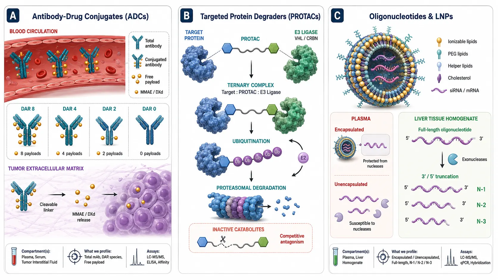 Figure 2: Multi-Component Bioanalytical Profiling Across Emerging Drug Modalities
Biological and Clinical Consequences of Relying on Single-Concentration Data
Relying on single-concentration bioanalytical methods for complex modalities introduces severe risks during
preclinical lead optimization and IND-enabling safety studies. Misidentifying the active driver of efficacy or
toxicity leads to erroneous SAR (structure-activity relationship) conclusions, inaccurate human dose predictions, and
failed clinical translation.
1. Masking Deconjugation Kinetics and Off-Target Toxicity
Consider an ADC candidate evaluated in a 14-day repeat-dose rodent toxicity study. If the bioanalytical assay
quantifies only "total antibody" via ligand-binding assay (LBA), the observed PK curve may exhibit an acceptable
half-life of 6 days with linear exposure scaling. However, if 40% of the payload is released into circulation within
the first 24 hours due to plasma esterase cleavage of the linker, the animal experiences severe bone marrow
suppression and liver toxicity. Without measuring free payload concentrations, toxicologists cannot attribute the
toxicity to the payload versus the antibody, potentially abandoning a promising target or advancing an unsafe linker
chemistry into human trials.
2. Distorted PK/PD Modeling and Exposure-Response Relationships
In non-compartmental analysis (NCA) and mechanistic PK/PD modeling, parameters such as clearance (CL), volume of
distribution (Vss), and area under the curve (AUC) are directly derived from analyte concentration. For a PROTAC
molecule, if an active metabolite is formed that retains target affinity but loses E3 ligase binding, measuring only
the parent drug concentration results in a severe mismatch between measured AUC and observed target degradation.
Incorporating simultaneous
parent and metabolite quantification services ensures that exposure metrics accurately reflect all
pharmacologically active species circulating in systemic and target tissue compartments.
Furthermore, mechanistic physiologically-based pharmacokinetic (PBPK) models for complex modalities rely heavily on
accurate multi-component inputs to simulate organ-level drug distribution and cleavage rates. Feeding a PBPK model
with only total drug concentration input leads to gross overestimation of active drug delivered to target tissues and
underestimates off-target organ exposure. Multi-component bioanalytical data provides the necessary granular input
parameters (unbound tissue partition coefficients, cleavage rate constants, and clearance values for each component)
to build predictive PBPK models that successfully translate preclinical findings to human trials.
3. Tissue Accumulation vs. Plasma Clearance Disconnects
Plasma concentration profiles frequently fail to mirror tissue exposure for novel modalities, particularly for LNPs
and GalNAc-conjugated oligonucleotides that preferentially accumulate in the liver, kidney, or tumor tissues. Rapid
plasma clearance does not necessarily imply drug elimination; it often reflects rapid tissue uptake and sequestration.
Multi-component analysis applied to tissue and cell lysate drug
quantification resolves the true tissue-to-plasma partition coefficients (Kp) for parent molecules, intra-tissue
released payloads, and localized catabolites.
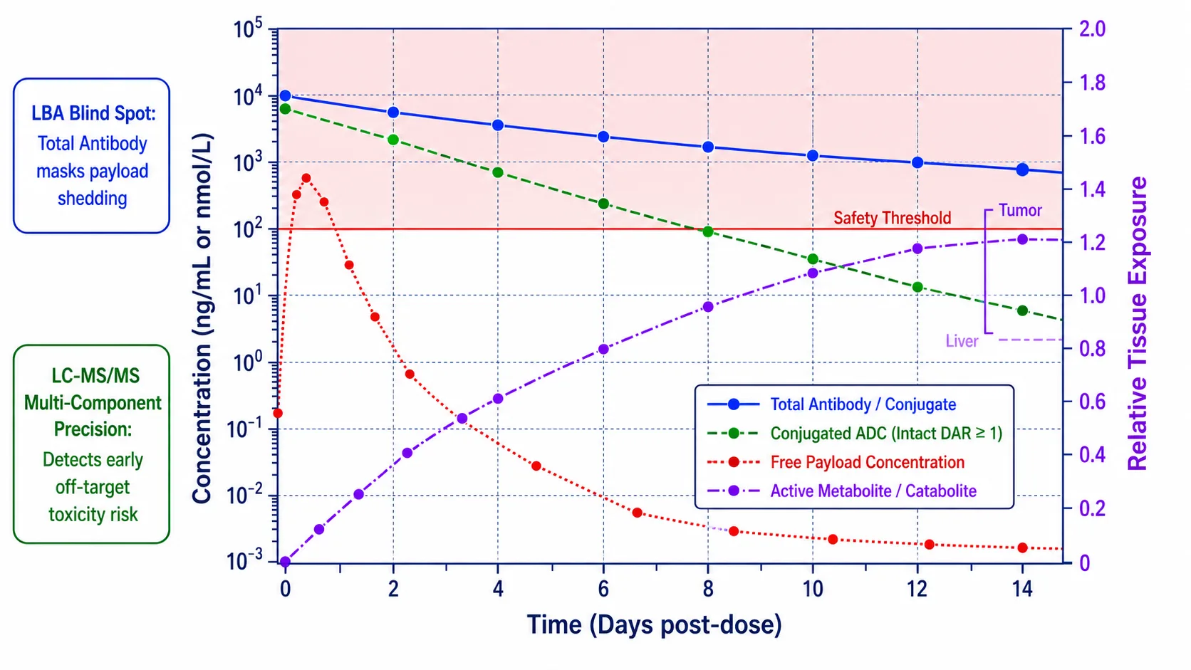 Figure 3: Pharmacokinetic Exposure Divergence: Total Conjugate vs. Free Payload and Active Catabolites
Advanced Bioanalytical Technologies and Workflows for Multi-Component Analysis
Achieving simultaneous, highly sensitive quantification of diverse analytes in a single biological sample requires
sophisticated hybrid instrumentation, tailored sample preparation strategies, and rigorous chromatographic separation.
1. Hybrid LBA-LC-MS/MS Platforms
Ligand-binding assays (LBA) provide exceptional sensitivity but lack molecular specificity, whereas LC-MS/MS provides
unambiguous mass specificity but can suffer from matrix interferences when analyzing intact biotherapeutics. Hybrid
LBA-LC-MS/MS bridges this gap. For ADCs and bispecifics, biotinylated target antigens or anti-human Fc antibodies are
immobilized on magnetic beads to selectively capture the therapeutic protein from plasma. Following washing, the
captured protein is subjected to either: (a) intact/middle-down mass spectrometry to evaluate DAR distribution, or (b)
controlled trypsin digestion followed by LC-MS/MS quantification of unique surrogate peptides and payload fragments.
Our high-sensitivity
multiplex quantification services leverage hybrid LBA-LC-MS/MS to achieve sub-ng/mL sensitivity across complex
bioanalytical panels.
2. Multi-Stage Sample Preparation: Dual Extraction Workflows
Multi-component panels often require measuring analytes with radically different physicochemical properties from the
same sample aliquot — for example, a 150 kDa intact antibody alongside a 700 Da hydrophobic payload (logP > 3).
Standard protein precipitation (PPT) is inadequate because it precipitates the antibody while co-extracting high
levels of matrix phospholipids that cause severe electrospray ion suppression. Advanced workflows employ dual-step
extraction: protein precipitation with organic solvent to isolate and re-dissolve small-molecule payloads/metabolites,
combined with sample preparation
method development services utilizing solid-phase extraction (SPE) or supported liquid extraction (SLE) to
achieve > 95% phospholipid removal prior to LC-MS/MS analysis.
3. High-Resolution LC-MS/MS and Multiplexed MRM/PRM
Triple quadrupole mass spectrometers operated in Multiple Reaction Monitoring (MRM) mode remain the workhorse for
small-molecule and peptide quantification due to their wide dynamic range (4–5 orders of magnitude) and high duty
cycle. When monitoring 20–50 transition channels simultaneously for multi-component panels, polarity switching and
dynamic MRM (retention time window triggering) ensure adequate dwell time (> 20 ms per transition) and sufficient data
points (> 15 points across a 3-second chromatographic peak). For ultra-complex catabolite screening and intact protein
mass resolution, Orbitrap or Q-TOF high-resolution mass spectrometry (HRMS) operating in parallel reaction monitoring
(PRM) or full-scan MS1 mode allows post-acquisition extraction of target mass channels with sub-5 ppm mass accuracy.
4. Microsampling Integration for Serial Rodent Pharmacokinetics
In preclinical discovery PK studies, obtaining full pharmacokinetic profiles from individual rodents has historically
been hampered by sample volume limitations (requiring composite sampling across multiple animals). Capillary
microsampling (CMS) and Volumetric Absorptive Microsampling (VAMS) collect 10–30 µL of whole blood per time point,
enabling full serial PK profiling in a single mouse or rat. Integrating microsampling with high-sensitivity
multi-component LC-MS/MS methods allows researchers to quantify parent drugs, free payloads, and active catabolites
from micro-volume samples while reducing animal usage by up to 70%. Supported by our microsampling bioanalysis
services, sponsors achieve high-throughput serial PK data without compromising sensitivity or analytical precision.
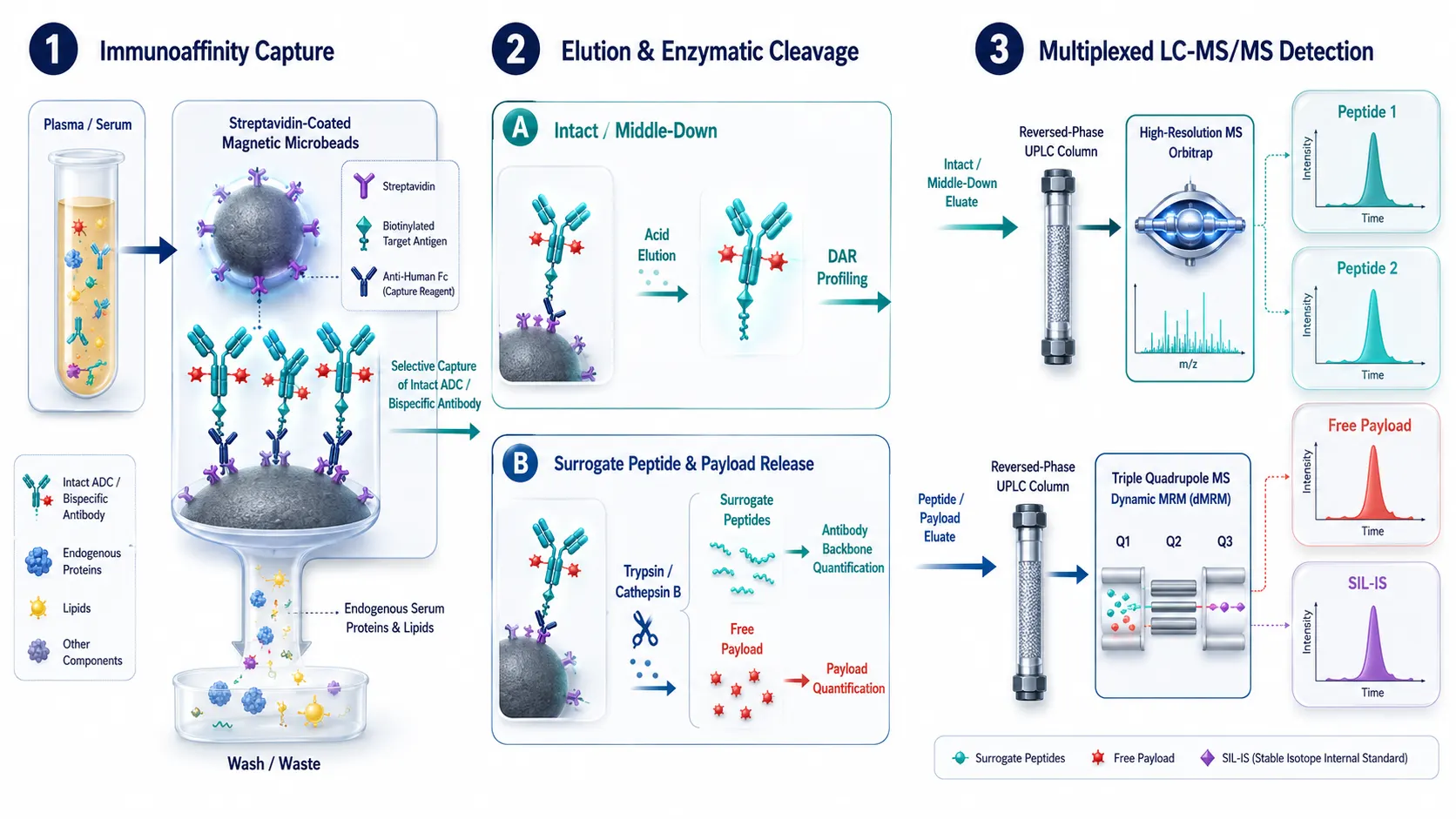 Figure 4: Hybrid LBA-LC-MS/MS Integrated Workflow for Multi-Component Quantification
Regulatory Compliance & Method Validation Under ICH M10 Standards
Bioanalytical methods supporting IND-enabling toxicology studies and clinical trials must undergo rigorous validation
in accordance with the ICH M10 global guideline. Multi-component assays introduce unique validation considerations
compared to single-analyte methods, as validation criteria must be independently demonstrated for every analyte within
the panel.
1. Matrix Factor & Ion Suppression Across Multiple Analytes
ICH M10 mandates the evaluation of matrix effects in at least six individual matrix lots. For multi-component panels,
the IS-normalized matrix factor CV must be ≤ 15% across all matrix lots for each individual analyte. Because
co-extracted matrix phospholipids elute across specific chromatographic retention windows, analytes in the panel with
differing retention times may experience distinct degrees of suppression. Chromatographic conditions must be optimized
to ensure that no analyte elutes within major phospholipid suppression zones.
2. Stability and Co-Formulated Analyte Interferences
Stability evaluation for multi-component panels must account for potential inter-analyte interconversion during
sample processing and storage. For instance, in an ADC panel, Bench-top stability must demonstrate that conjugated
payload does not undergo non-enzymatic deconjugation into free payload in the autosampler or during freeze-thaw
cycles. Supported by our short-term and long-term stability study services and forced degradation profiling, sponsors
receive fully validated stability datasets covering freeze-thaw, bench-top, autosampler, and long-term storage
conditions.
3. Incurred Sample Reanalysis (ISR) Strategy
Incurred Sample Reanalysis (ISR) verifies the repeatability of the bioanalytical method using actual study samples
rather than spiked QCs. For multi-component assays, ISR acceptance criteria (at least 67% of reanalyzed samples within
±20% of the original concentration) must be met independently for every quantified component in the panel. Passing ISR
across all analytes confirms that sample handling, protein binding, and in vivo matrix components do not compromise
bioanalytical accuracy.
Decision Matrix: Single-Analyte vs. Multi-Component Bioanalysis
Selecting the appropriate bioanalytical strategy depends on the therapeutic modality, development stage, and
regulatory context. The following decision matrix compares single-analyte and multi-component bioanalytical approaches
across key operational parameters:
| Parameter |
Single-Analyte Bioanalysis |
Multi-Component Bioanalysis |
| Primary Scope |
Single parent drug or total antibody concentration |
Parent drug, conjugated species, free payload, catabolites, and biomarkers |
| Applicable Modalities |
Traditional small molecules, simple mAb therapeutics |
ADCs, PROTACs/TPDs, bispecifics, oligonucleotides, LNPs |
| Analytical Specificity |
Moderate; vulnerable to co-eluting active/inactive metabolites |
High; unambiguous mass-based resolution of parent and all catabolites |
| PK/PD Insight Level |
Basic exposure metrics (Cmax, AUC, t1/2); fails to capture deconjugation |
Mechanistic exposure-safety-efficacy profiling; captures biotransformation kinetics |
| Method Development Complexity |
Low to Moderate; single extraction and chromatographic optimization |
High; requires dual-extraction, multiplex MRM/PRM, and stable isotope IS |
| Regulatory Alignment (ICH M10) |
Sufficient for traditional discovery PK screening |
Recommended for IND-enabling safety, GLP tox, and clinical translation of complex modalities |
Implementing multi-component bioanalysis early in drug discovery avoids costly late-stage failures. Supported by our
custom LC-MS/MS method development for novel chemical entities and rigorous full and partial method validation
services under ISO/IEC 17025 accreditation, sponsors receive regulatory-ready bioanalytical packages tailored to the
unique biotransformation profiles of their lead candidates.
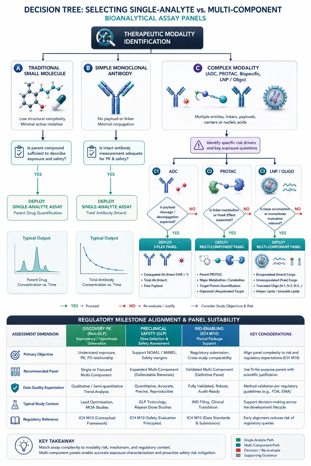 Figure 5: Decision Tree for Deploying Multi-Component Assay Panels in Preclinical DMPK
Frequently Asked Questions
Why is single-analyte quantification insufficient for Antibody-Drug Conjugates (ADCs)?
ADCs undergo continuous systemic biotransformation in vivo, including payload deconjugation, antibody
degradation, and catabolite formation. Measuring only total antibody fails to detect premature payload shedding,
which drives systemic toxicity. Measuring only free payload fails to track tumor-targeted delivery. A complete
ADC profile requires simultaneous quantification of conjugated antibody, total antibody, and free
payload/catabolites.
What key analytes must be quantified in a PROTAC multi-component panel?
A comprehensive PROTAC bioanalytical panel typically quantifies intact parent PROTAC, major cleavage products
(e.g., monovalent warhead or E3 ligase binder fragments that can cause competitive antagonism), and
pharmacodynamic markers of target protein degradation. Tracking parent-to-metabolite ratios is essential to
distinguish clearance from metabolic inactivation.
How does hybrid LBA-LC-MS/MS overcome matrix interference in tissue bioanalysis?
Hybrid LBA-LC-MS/MS uses specific antibody or antigen capture (LBA step) to selectively isolate the target
molecule from dense tissue homogenate matrix components, eliminating > 99% of interfering proteins and lipids.
Subsequent enzymatic digestion and LC-MS/MS analysis provide precise, mass-specific quantification of surrogate
peptides or payloads with minimal matrix suppression.
What sample volume is required for multi-component LC-MS/MS panels in rodent PK studies?
With modern high-sensitivity triple quadrupole mass spectrometers, multi-component panels can be routinely
executed using 25–50 µL of plasma or serum per sample. For non-terminal rodent serial sampling, microsampling
techniques (such as VAMS or capillary microsampling) enable reliable multi-component quantification from 10–20
µL of whole blood.
How are internal standards chosen for multi-component bioanalytical assays?
Ideally, stable isotope-labeled internal standards (SIL-IS, such as 13C- or 15N-labeled analogs) should be
incorporated for each primary analyte in the panel to correct for matrix effects and extraction recovery
variations. When SIL-IS is unavailable for novel metabolites or payloads, closely matched structural analogs
with validated matrix-tracking performance are utilized.
Do multi-component bioanalytical methods comply with ICH M10 regulatory guidance?
Yes. Multi-component methods developed for IND-enabling GLP toxicology and clinical studies undergo full
validation per ICH M10 guidelines, evaluating selectivity, calibration curve linearity, accuracy, precision,
matrix factor variability across ≥ 6 independent lots, carryover, and stability under all processing and storage
conditions for each analyte in the panel.
How is Incurred Sample Reanalysis (ISR) conducted for multi-component panels?
ISR is performed by re-analyzing a subset of study samples (typically 10% of the first 1000 samples and 5% of
remaining samples). For multi-component panels, the percent difference between original and repeat values must
be within ±20% for at least 67% of re-analyzed samples independently for each analyte in the panel.
How do you prevent MRM channel crosstalk and carryover in multi-component panels?
Crosstalk occurs when structurally similar analytes or metabolites yield overlapping precursor/product ion
pairs. To prevent crosstalk, chromatographic baseline resolution (> 0.3 min separation) is established between
co-monitored analytes, and inter-channel delay times are optimized in the collision cell. Carryover is minimized
using needle-wash solvent optimization and gradient flushing steps.
References
- ICH Harmonised Guideline. Bioanalytical Method Validation and Study Sample
Analysis M10. International Council for Harmonisation of Technical Requirements for Pharmaceuticals for
Human Use; 2022.
- Gorovits B, Alley SC,
Bilic S, et al. Bioanalysis of antibody-drug conjugates: American Association of Pharmaceutical Scientists
recommendation. Bioanalysis. 2013;5(9):1057-1072.
- Pike A, Bilsland B,
Harrington EA, et al. Optimizing PROTAC degradation kinetics and exposure for preclinical drug discovery.
Journal of Medicinal Chemistry. 2020;63(9):4884-4897.
- Xu RN, Fan L, Rieser MJ,
El-Shourbagy TA. Recent advances in hybrid LBA-LC-MS/MS bioanalysis of biotherapeutics and biomarkers.
Journal of Chromatography B. 2019;1118:122-135.
- Bioanalytical assay strategies for
the development of antibody-drug conjugate biotherapeutics. Bioanalysis. 2013;5(2):201-226.
- Jemal M, Ouyang Z. Bioanalysis of
complex modalities by LC-MS/MS: Strategies for sample preparation and chromatographic resolution.
Biomedical Chromatography. 2020;34(3):e4782.
- Korfmacher WA. Principles
and applications of LC-MS in novel drug discovery and DMPK characterization. Drug Discovery Today.
2005;10(20):1357-1367.
- Hendrikx JJMA, Rosing H, Schinkel AH,
Beijnen JH. Quantification of intact biotherapeutics and free payloads by multiplexed LC-MS/MS.
Rapid Communications in Mass Spectrometry. 2020;34(12):e8821.
- Tiller PR, Romanyshyn LA.
Liquid chromatography-tandem mass spectrometry in drug discovery and development: Multi-component assay
design. Journal of Pharmaceutical and Biomedical Analysis. 2021;198:114120.
- Quantification of mRNA in
Lipid Nanoparticles Using Mass Spectrometry. Analytical Chemistry. 2024;96(3):3658-3665.
Related Services


