Why CYP-Mediated DDI Matters: The Clinical and Regulatory Stakes
Cytochrome P450 (CYP) enzymes metabolize approximately 75% of all marketed drugs, and CYP3A4 alone accounts for roughly 50% of that share. When two drugs that share a CYP-mediated clearance pathway are co-administered, one drug can alter the exposure of the other — sometimes catastrophically. The clinical pharmacology literature is replete with cautionary cases: terfenadine withdrawn in 1998 after CYP3A4 inhibition by ketoconazole and erythromycin produced life-threatening QTc prolongation; cerivastatin withdrawn in 2001 after gemfibrozil's CYP2C8 inhibition caused rhabdomyolysis; mibefradil withdrawn within a year of launch after its potent CYP3A4 TDI precipitated fatal DDIs with beta-blockers, digoxin, and verapamil. Each of these cases was preventable with the in vitro CYP DDI assays that are now standard in drug development — but those assays did not systematically exist at the time. CYP-mediated drug-drug interaction assessment is now a non-negotiable regulatory gate that every small-molecule drug candidate must pass before entering Phase II.
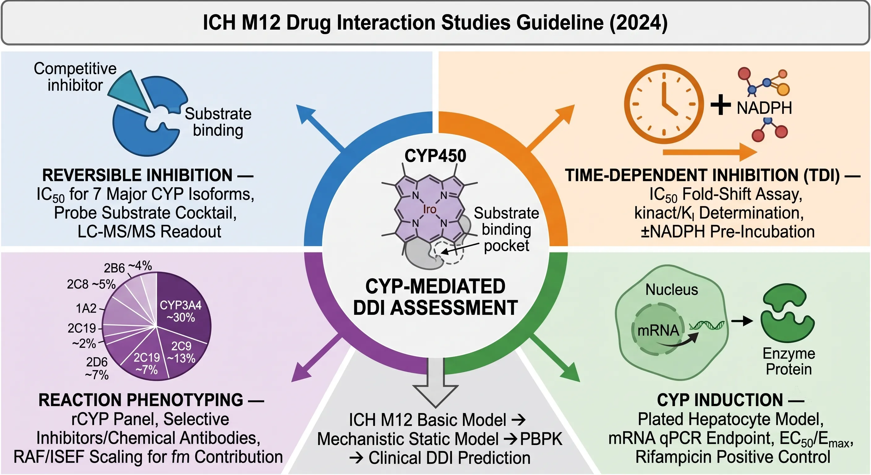 Figure 1: The CYP-Mediated DDI Assessment Workflow — Reversible Inhibition, TDI, Induction, and Phenotyping per ICH M12
The publication of ICH M12 (Drug Interaction Studies, adopted May 2024, effective November 2024) is the single most important regulatory event in DDI assessment in two decades. Prior to ICH M12, DDI guidances operated in regional silos — FDA 2020, EMA 2013, PMDA 2018 — each with different CYP isoform panels, different cutoff criteria, and different expectations for TDI and induction study design. ICH M12 harmonizes these into a single globally acceptable framework, and in doing so introduces substantive scientific changes: the TDI clinical cutoff scaling factor reduced from 50× to 5× Cmax, mRNA designated as the preferred primary CYP induction endpoint, and the Basic Model → Mechanistic Static Model (MSM) → PBPK decision tree codified as the standard approach for translating in vitro data into clinical DDI predictions. Every drug development team submitting to global regulatory agencies must now align their CYP DDI assessment strategy with ICH M12.
The stakes of getting CYP DDI assessment wrong extend beyond individual patients to entire development programs. A missed CYP2D6 TDI signal at IND can become a clinical hold when a Phase IIb study enrolls a patient on paroxetine who experiences a 5-fold increase in exposure to the investigational drug. Conversely, an overestimated CYP3A4 induction risk can lead to an unnecessary clinical DDI study costing $500,000-$1.5 million and delaying the program by 6-12 months. The challenge is not whether to evaluate CYP-mediated DDI — that question is settled — but how to evaluate it efficiently, interpret the data correctly per ICH M12, and make informed decisions about which signals warrant clinical follow-up and which can be dismissed by the tiered in vitro-to-clinical prediction framework.
The ICH M12 Framework for CYP DDI: When to Test and What the Guidance Requires
ICH M12 organizes CYP-mediated DDI assessment into four sequential experimental domains — reversible inhibition, time-dependent inhibition (TDI), induction, and reaction phenotyping — with each domain feeding data into a unified clinical risk prediction algorithm. The framework is not a checklist where every assay is performed on every compound. Instead, it is a decision architecture where results from earlier steps determine which subsequent steps are necessary and at what level of resolution.
Reversible inhibition is the starting point and the most broadly applicable assay. Every small-molecule drug candidate entering clinical development is expected to have IC50 values determined against the seven major CYP isoforms: CYP1A2, CYP2B6, CYP2C8, CYP2C9, CYP2C19, CYP2D6, and CYP3A4. The IC50 experiment uses human liver microsomes (HLM) as the enzyme source, a cocktail of isoform-selective probe substrates, and LC-MS/MS quantification of the metabolite products. If the Basic Model cutoff (Cmax,u/Ki,u < 0.02) is met for all seven isoforms, the reversible inhibition assessment is complete and no clinical DDI study is triggered by this mechanism. If the cutoff is exceeded for any isoform, the risk is further refined through the Mechanistic Static Model.
Time-dependent inhibition (TDI) is evaluated when the chemical structure suggests potential for mechanism-based inactivation — structural alerts include terminal alkyne, furan, thiophene, methylenedioxyphenyl, and alkylamine functional groups — or when the compound is intended for chronic administration where cumulative enzyme inactivation is clinically relevant. The TDI assessment uses a ±NADPH pre-incubation IC50 fold-shift assay. A ≥1.5-fold decrease in IC50 in the +NADPH arm relative to the −NADPH arm triggers a full kinact/KI determination. The clinical relevance of TDI is evaluated through the (kobs+kdeg)/kdeg ratio: a value < 1.25 excludes TDI concern per ICH M12.
CYP induction evaluation is required when: (a) the drug is intended for chronic administration (≥7 days), (b) it shows nuclear receptor activation (PXR, CAR, AhR) in reporter gene assays, or (c) it belongs to a chemical class with known induction liability. The standard model uses three individual lots of plated human hepatocytes treated with the test compound for 48-72 hours, with CYP1A2, CYP2B6, and CYP3A4 mRNA measured by qPCR as the primary endpoint. A concentration-dependent increase ≥2-fold at concentrations ≤50× unbound Cmax is considered induction-positive. Secondary evaluation of CYP2C8, CYP2C9, and CYP2C19 induction is only required if CYP3A4 is induced, since these isoforms share the PXR-mediated induction mechanism.
Reaction phenotyping identifies which CYP isoform(s) are responsible for metabolizing the drug candidate and quantifies their fractional contribution (fm) to total clearance. It is required when metabolism accounts for >25% of total clearance, and it directly informs the MSM DDI prediction by providing the fm parameter — without fm, the MSM cannot calculate the predicted AUC ratio because the fraction of clearance susceptible to inhibition or induction is unknown.
The operational integration of these four domains into the transporter-mediated drug-drug interaction assessment that completes the ICH M12 evaluation is the subject of the complementary article in this series. Together, CYP and transporter DDI assessment comprise the full in vitro DDI package required for an IND or NDA submission under ICH M12.
Reversible CYP Inhibition: IC50 Determination Across Seven Major Isoforms
The reversible CYP inhibition assay answers a deceptively simple question: does the test compound compete with the probe substrate for the CYP active site? The experimental design places the drug candidate — the potential perpetrator — in competition with an isoform-specific probe substrate in HLM, and measures whether the rate of probe metabolite formation decreases in a concentration-dependent manner. The result is an IC50: the perpetrator concentration that reduces metabolite formation by 50%. If the IC50 is high relative to the expected clinical concentration at the enzyme active site (unbound Cmax at the liver inlet), the compound is a weak inhibitor and the DDI risk is low. If the IC50 is close to or below the clinical concentration, the compound is a potent inhibitor and a clinical DDI study may be warranted.
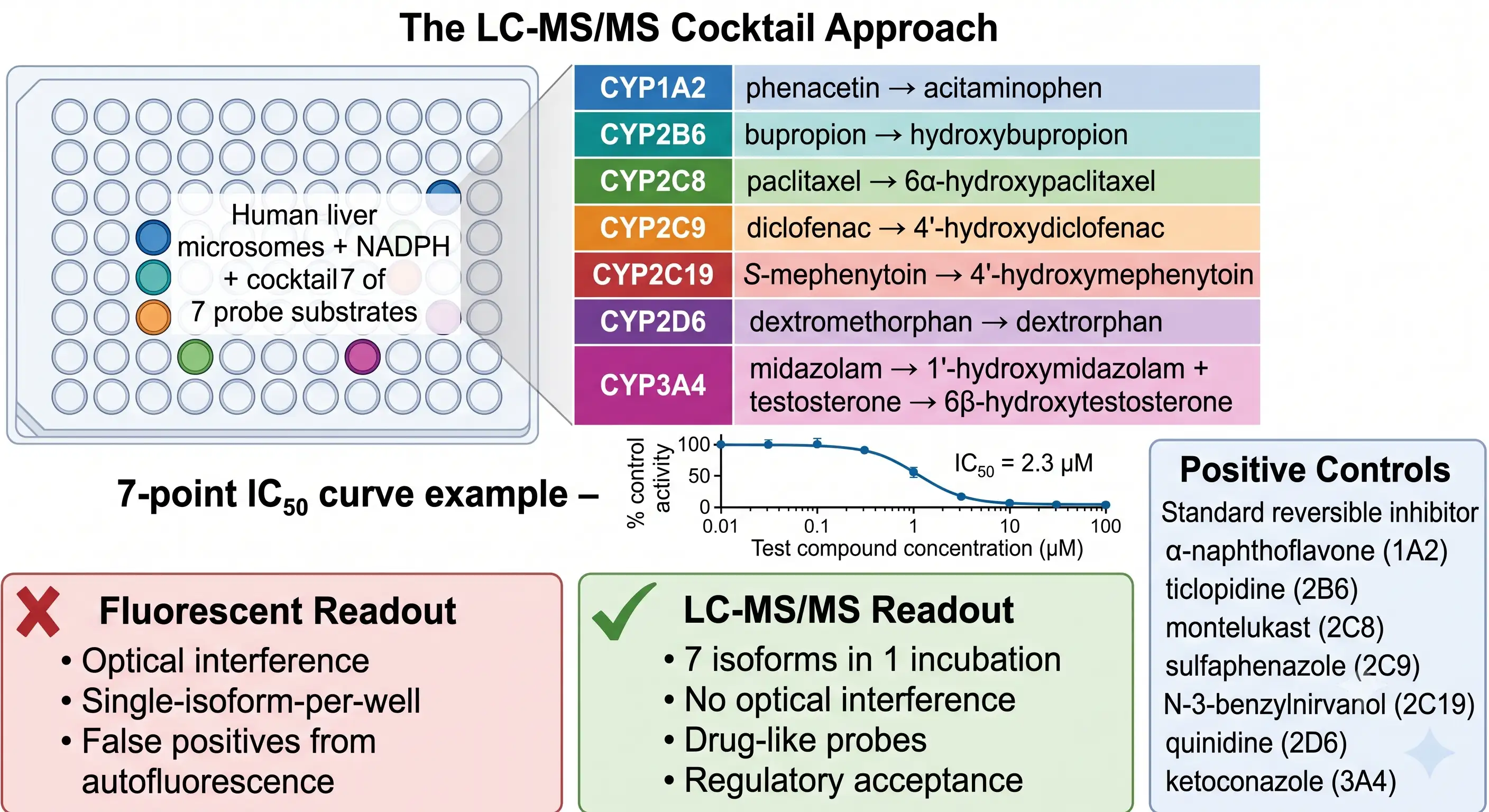 Figure 2: Reversible CYP Inhibition — The Seven-Isoform IC50 Determination Panel
The modern approach uses an LC-MS/MS probe substrate cocktail — all seven isoform-selective probe substrates are combined in a single incubation well with HLM, NADPH, and the test compound at multiple concentrations. After incubation, all seven metabolite products are quantified simultaneously by LC-MS/MS in a single analytical run. This cocktail approach replaced the earlier fluorescent-based method, which had three fundamental limitations: (1) optical interference — test compounds that fluoresce or quench fluorescence produce false-positive or false-negative IC50 values; (2) one-isoform-per-well format requiring seven separate incubations and analyses; and (3) non-selective fluorescent probes that lacked isoform specificity at higher concentrations. The LC-MS/MS cocktail method with drug-like probe substrates — phenacetin (1A2), bupropion (2B6), paclitaxel (2C8), diclofenac (2C9), S-mephenytoin (2C19), dextromethorphan (2D6), and midazolam + testosterone (3A4) — is now the regulatory standard endorsed by ICH M12.
The experimental protocol for an IC50 determination is straightforward but sensitive to several parameters that, if not controlled, produce misleading results. Substrate concentration must be at or below the Km for each probe substrate — if the probe substrate concentration exceeds Km, the inhibitor is competing against excess substrate, and the observed IC50 overestimates the true inhibitory potency. The Cheng-Prusoff equation (Ki = IC50 / [1 + S/Km]) corrects for substrate competition and converts IC50 to the substrate-independent inhibition constant Ki, which is the parameter used in the Basic Model. Microsomal protein concentration must be kept low (typically 0.1-0.5 mg/mL) to minimize non-specific binding of lipophilic test compounds to microsomal phospholipids — a compound that partitions extensively into microsomal membranes has a lower free concentration in the aqueous phase than the nominal concentration, and the apparent IC50 is artificially elevated. Organic solvent concentration (typically DMSO or acetonitrile for test compound stocks) must be kept below 0.1-1% v/v, as organic solvents inhibit CYP activity in an isoform-dependent manner: CYP3A4 is particularly sensitive to DMSO, with 1% DMSO producing up to 20% inhibition in some HLMs.
Positive control inhibitors are run in parallel with every experiment to qualify the HLM batch and confirm assay sensitivity: α-naphthoflavone (CYP1A2), ticlopidine (CYP2B6), montelukast (CYP2C8), sulfaphenazole (CYP2C9), N-3-benzylnirvanol (CYP2C19), quinidine (CYP2D6), and ketoconazole (CYP3A4). If the IC50 of the positive control deviates by more than 2-fold from the historical laboratory mean, the HLM batch activity is suspect and the experiment should be repeated with a fresh batch. The intersection of reversible CYP inhibition data with in vitro compound stability profiling is operationally important: compounds with poor microsomal stability (high intrinsic clearance) may be extensively metabolized during the IC50 incubation, reducing the effective inhibitor concentration and producing a falsely elevated IC50. Metabolic stability data should be reviewed alongside IC50 data to identify this confound.
Time-Dependent Inhibition (TDI): kinact/KI, IC50 Shift, and Mechanism-Based Inactivation
Time-dependent inhibition differs from reversible inhibition in mechanism, detection, and clinical consequence. A reversible inhibitor binds non-covalently and its effect dissipates as the inhibitor is cleared from the body — the maximum DDI risk coincides with the Cmax of the perpetrator. A TDI compound is metabolically activated by the CYP enzyme to a reactive intermediate that covalently modifies the enzyme, permanently inactivating it. The inactivated enzyme is gone — it must be replaced by de novo CYP protein synthesis, a process with a half-life of 36 hours (CYP3A4) to 140 hours (CYP2E1). During that period, all substrates of the inactivated CYP isoform are subject to reduced clearance regardless of whether the perpetrator is still present in the circulation. TDI DDIs are typically larger in magnitude, longer in duration, and less predictable from Cmax timing than reversible inhibition DDIs.
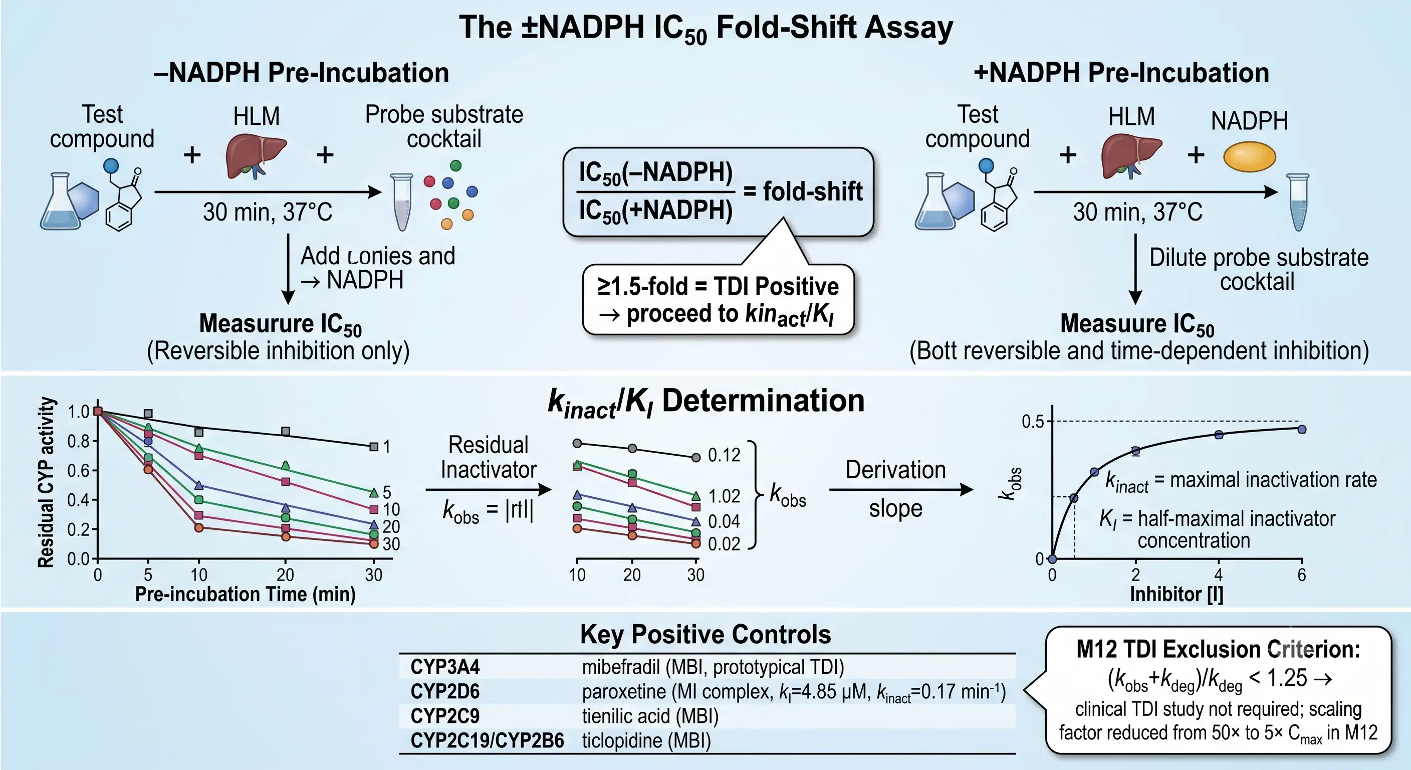 Figure 3: Time-Dependent CYP Inhibition (TDI) — The IC50 Fold-Shift Assay and kinact/KI Determination
The primary TDI screen is the ±NADPH IC50 fold-shift assay (also called the IC50 shift or dilution assay). Two parallel pre-incubations are set up, each containing test compound (at multiple concentrations) and HLM. The −NADPH arm receives buffer only; the +NADPH arm receives NADPH to initiate CYP catalytic activity. After a 30-minute pre-incubation at 37°C, both arms are diluted (typically 10-fold) into a solution containing the probe substrate cocktail and NADPH, and the residual CYP activity is measured. If the test compound is a TDI, the +NADPH arm will show a lower IC50 than the −NADPH arm because the CYP enzyme has been partially inactivated during the pre-incubation. An IC50 shift ≥1.5-fold (IC50(−NADPH)/IC50(+NADPH) ≥ 1.5) is considered TDI-positive and triggers a full kinact/KI determination. The dilution step is critical: without it, the reversible inhibition component of the total inhibition in the +NADPH arm cannot be distinguished from the irreversible TDI component. The 10-fold dilution reduces the concentration of both test compound and probe substrate, effectively eliminating the reversible component while leaving the irreversible enzyme inactivation intact.
For compounds that are TDI-positive by the fold-shift screen, the kinact/KI determination provides the kinetic parameters needed for clinical DDI prediction. The experiment measures residual CYP activity as a function of pre-incubation time (typically 0, 5, 10, 20, and 30 minutes) at multiple inhibitor concentrations. For each inhibitor concentration, the natural log of residual activity vs time yields a straight line with slope = −kobs. The kobs values are then plotted against inhibitor concentration, and the resulting hyperbolic plot is fitted to kobs = kinact × [I]/(KI + [I]) to yield kinact (the maximal inactivation rate constant at saturating inhibitor concentration) and KI (the inhibitor concentration producing half-maximal inactivation rate). A compound with a low KI and a high kinact is the most dangerous TDI perpetrator — it inactivates the enzyme efficiently at low clinical concentrations.
The key ICH M12 change for TDI assessment is the reduction of the clinical cutoff scaling factor from 50× to 5× Cmax. Under the FDA 2020 guidance, the Basic Model TDI equation (kobs+kdeg)/kdeg used a predicted unbound liver inlet concentration of 50 × Cmax,u — essentially, every TDI-positive compound was flagged for clinical study because the hugely conservative concentration assumption overwhelmed the kinetic data. ICH M12 replaces the 50× factor with 5×, reflecting a more physiologically relevant estimate of unbound liver concentration. The consequence: fewer false-positive TDI flags, fewer unnecessary clinical DDI studies, and a stronger reliance on the kinact/KI data to discriminate between clinically relevant and clinically irrelevant TDI.
Positive controls for TDI assays are mechanism-specific. Mibefradil (withdrawn TDI-positive calcium channel blocker) is the prototypical CYP3A4 mechanism-based inactivator, producing a >10-fold IC50 shift. Paroxetine is a CYP2D6 TDI-positive control (KI = 4.85 µM, kinact = 0.17 min⁻¹) that forms a quasi-irreversible metabolite-intermediate (MI) complex rather than covalent heme adduction — a mechanistically distinct form of TDI that is NADPH-dependent but reversible under certain conditions. Tienilic acid (CYP2C9) and ticlopidine (CYP2C19/CYP2B6) are mechanism-based inactivators with well-characterized kinetics. The positive control should be selected to match the suspected TDI mechanism — MI complex formation vs covalent heme modification vs apoprotein alkylation — because the clinical consequences and the (kobs+kdeg)/kdeg cutoff interpretation differ.
CYP Induction: Plated Hepatocyte Model, mRNA vs Activity Endpoints, and EC50 Determination
CYP induction is the counterpoint to CYP inhibition: instead of reducing enzyme activity, the perpetrator drug increases it by activating nuclear receptors (PXR for CYP3A4/CYP2C, CAR for CYP2B6, AhR for CYP1A2) that upregulate CYP gene transcription, increasing the total enzyme pool. The clinical consequence of CYP induction is reduced exposure of the victim drug, potentially to subtherapeutic levels. Rifampicin, the prototypical broad-spectrum inducer, reduces the AUC of oral midazolam (a CYP3A4 probe substrate) by >90% — a magnitude of effect that can render a co-administered contraceptive, anticoagulant, or antiretroviral completely ineffective. Induction is a chronic-dosing phenomenon: enzyme upregulation requires sustained nuclear receptor activation over days, and the full induction effect plateaus after approximately 5-7 days of daily dosing.
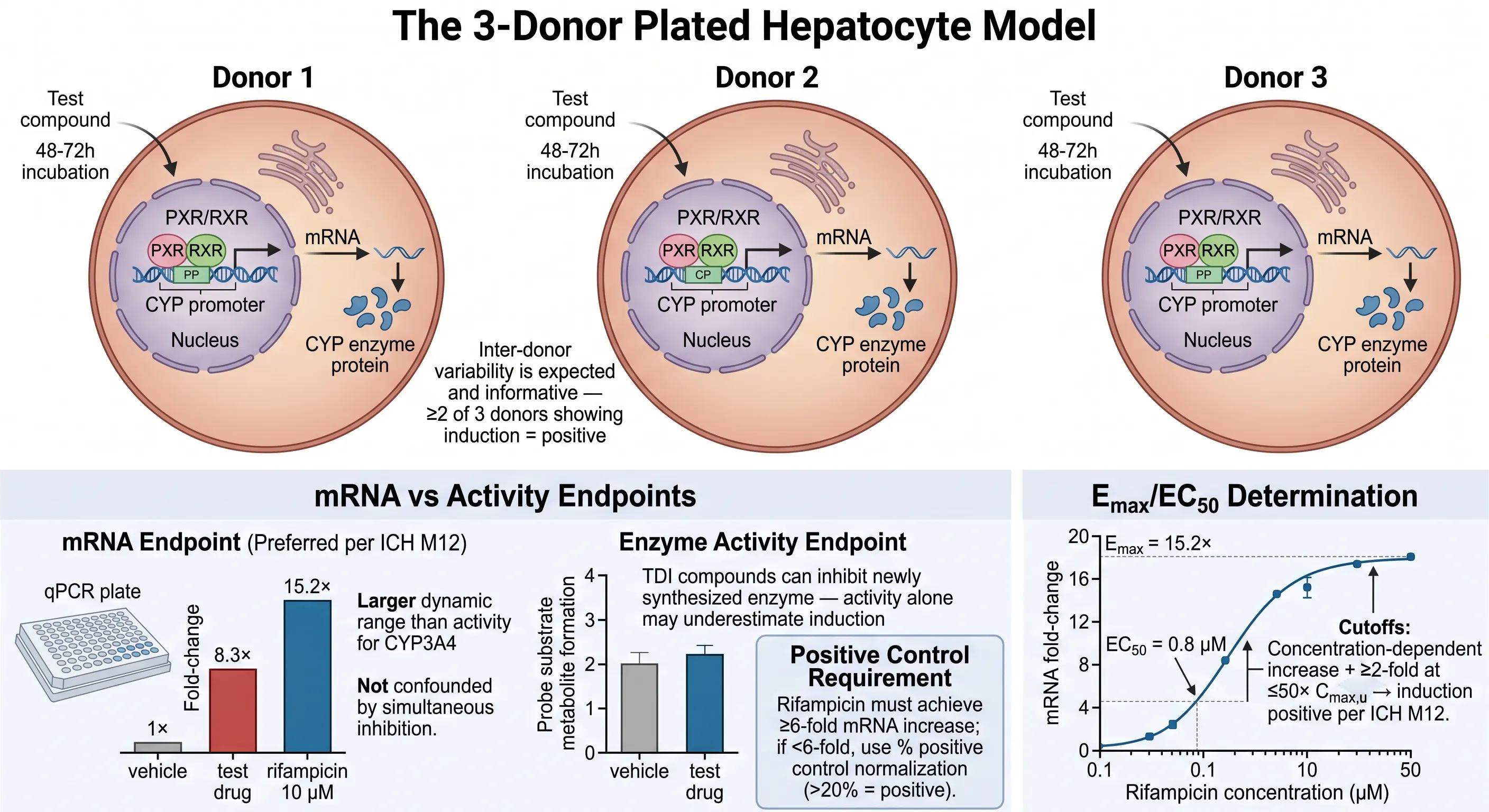 Figure 4: CYP Induction — Plated Hepatocyte Model, mRNA vs Activity Endpoints
The ICH M12-recommended induction model uses three individual lots of plated human hepatocytes, with the test compound added to culture medium for 48-72 hours — sufficient time for transcriptional activation, mRNA accumulation, and enzyme protein synthesis. The use of three donors captures inter-individual variability in PXR/CAR expression and responsiveness, which is substantial: CYP3A4 induction by rifampicin can range from 5-fold to 30-fold across individual hepatocyte donors. Per ICH M12, a positive induction finding requires ≥2 of 3 donors showing a concentration-dependent increase — one donor with an unusually responsive PXR phenotype should not drive a positive call, nor should two non-responsive donors mask a genuine induction liability.
mRNA (measured by qPCR or branched DNA) is the preferred primary endpoint per ICH M12, with two decisive advantages over enzyme activity. First, the dynamic range is substantially larger: a strong inducer like rifampicin produces 10-30-fold mRNA increases for CYP3A4, compared to 2-5-fold increases in testosterone 6β-hydroxylase activity. This compressed activity dynamic range reduces the statistical power to distinguish moderate inducers from non-inducers. Second, and mechanistically more important, mRNA measurement is not confounded by simultaneous CYP inhibition. A test compound that is both an inducer and an inhibitor — a common profile, since many CYP3A4 inducers are also CYP3A4 substrates and competitive inhibitors — can produce a false-negative result in an activity-based assay because the inhibitor effect during the terminal activity incubation partially or fully masks the increased enzyme pool. In the mRNA assay, cells are lysed for RNA extraction after the induction period; no enzyme activity incubation occurs, and the inhibitor never has the opportunity to interfere with the mRNA readout.
Enzyme activity remains valuable as a confirmatory secondary endpoint. A compound that produces ≥2-fold CYP3A4 mRNA increase but no corresponding activity increase is "mRNA-positive, activity-negative" — a pattern that suggests simultaneous inhibition and warrants follow-up investigation, such as a washout experiment where the test compound is removed before the activity assay, or an orthogonal induction endpoint (western blot for CYP3A4 protein). The ICH M12 induction-positive criterion is: concentration-dependent increase ≥2-fold at ≤50× unbound Cmax,u, with a clear concentration-response relationship and ≥2 of 3 donors responding. Rifampicin (10 µM) must produce ≥6-fold CYP3A4 mRNA increase as the positive control; if <6-fold, the hepatocyte batch has inadequate induction responsiveness and the experiment must be repeated.
The induction assessment is not complete without integrating the clinical context. CYP1A2 and CYP3A4 are the most clinically significant induction targets because they have the broadest substrate coverage. CYP2B6 induction is important for drugs metabolized by this isoform (efavirenz, bupropion, methadone). Induction of CYP2C8, CYP2C9, and CYP2C19 is evaluated as a secondary panel — only when CYP3A4 induction is positive — because these isoforms share the PXR regulatory mechanism. A compound that does not induce CYP3A4 is extremely unlikely to induce CYP2C9 or CYP2C19, and separate induction assessment is not warranted. The induction data feeds into the MSM and PBPK models as Emax (maximal fold-induction) and EC50 (concentration producing half-maximal induction), which together describe the full induction concentration-response relationship for quantitative DDI prediction.
Reaction Phenotyping: Identifying Which CYP Metabolizes Your Drug
Reaction phenotyping answers a question that is logically prior to inhibition and induction assessment: which CYP isoform is responsible for clearing the drug? The answer — expressed as fm,CYP3A4, fm,CYP2D6, etc. — determines which inhibition and induction findings are clinically relevant. If CYP3A4 contributes 80% of total clearance (fm,CYP3A4 = 0.8), a potent CYP3A4 inhibitor will produce a large AUC change, while a potent CYP2D6 inhibitor will produce a small or negligible change. Without reaction phenotyping data, the DDI risk of a perpetrator drug cannot be evaluated because the fraction of clearance at risk — the fm — is unknown. ICH M12 requires reaction phenotyping for any drug where metabolism accounts for >25% of total clearance and the metabolic pathway involves CYP enzymes.
The standard phenotyping strategy uses two complementary experimental approaches in human liver microsomes. The recombinant CYP (rCYP) panel — also called CYP mapping — incubates the test compound at a low, clinically relevant concentration with individual recombinant CYP enzymes (CYP1A2, 2B6, 2C8, 2C9, 2C19, 2D6, and 3A4) and measures the rate of substrate depletion or metabolite formation. The pattern of activity across isoforms identifies which CYPs are capable of metabolizing the drug, but not their quantitative contribution in the native HLM environment where CYP expression levels differ dramatically (CYP3A4 is the most abundant hepatic CYP, while CYP2D6 constitutes only ~2% of total CYP content). Selective chemical inhibitor or CYP-specific antibody inhibition in HLM provides the quantitative fm. If a selective CYP3A4 inhibitor (ketoconazole at 1 µM, or an anti-CYP3A4 monoclonal antibody) reduces the metabolism rate by 70% in pooled HLM, then CYP3A4 contributes approximately 70% to the hepatic clearance of that compound (fm,CYP3A4 = 0.7). The inhibitor concentration and isoform selectivity must be carefully validated: ketoconazole at concentrations >1 µM loses CYP3A4 selectivity and begins inhibiting CYP2C8 and CYP2C9.
The quantitative scaling from recombinant CYP data to native HLM uses Relative Activity Factor (RAF) or Intersystem Extrapolation Factor (ISEF). RAF is the ratio of the activity of a reference CYP isoform-selective reaction in HLM to the same reaction in the rCYP system. ISEF extends RAF by incorporating immunoquantified CYP abundance data, providing a more direct protein-to-activity correlation. The rCYP intrinsic clearance for each isoform is multiplied by the corresponding RAF/ISEF to estimate the contribution of that isoform to the total HLM intrinsic clearance, and fm = CLint,HLM,CYPx / CLint,HLM,total. An fm > 0.25 for a single CYP isoform flags that isoform as the major clearance pathway — a DDI involving that isoform is potentially clinically significant and must be evaluated through the MSM or PBPK prediction framework.
Reaction phenotyping intersects directly with drug metabolite identification by LC-MS in two ways. First, identifying the metabolite(s) produced from the phenotyping incubation confirms that the rCYP isoform is genuinely responsible for that specific metabolic pathway, rather than a non-specific loss of parent that could reflect binding or instability. Second, if the metabolite profile in the rCYP incubation matches the human in vivo metabolite profile, the in vitro phenotyping result is physiologically relevant. If the rCYP produces a metabolite not observed in vivo, or if the in vivo metabolite is not produced by any rCYP, the phenotyping is incomplete — non-CYP enzymes (FMOs, MAOs, AO, CES) or extrahepatic metabolism may be contributing to clearance in ways the rCYP panel cannot detect.
From In Vitro Data to Clinical DDI Prediction: Static Models, PBPK, and ICH M12 Cutoff Criteria
The end goal of CYP inhibition, TDI, and induction experiments is not the raw IC50, Ki, kinact/KI, or Emax/EC50 value — it is the predicted clinical AUC ratio (AUCR) for a co-administered victim drug. ICH M12 codifies a three-tiered decision framework for translating in vitro CYP data into clinical DDI predictions, with each tier applying progressively more complex and physiologically realistic modeling while filtering out compounds whose DDI risk can be confidently excluded by simpler methods.
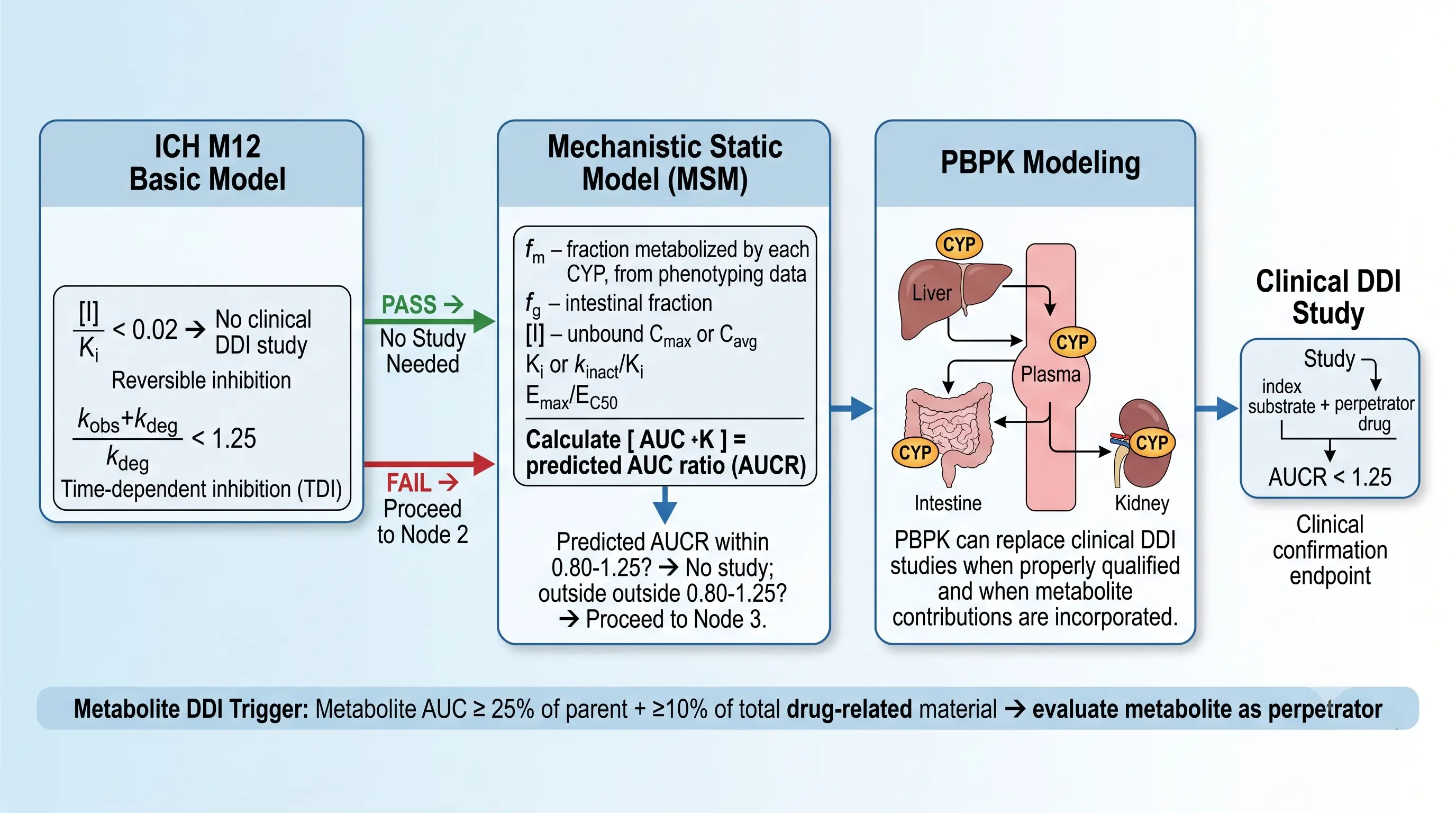 Figure 5: From In Vitro Data to Clinical DDI Prediction — Basic Model, Mechanistic Static Model, and PBPK Decision Tree
Tier 1 — ICH M12 Basic Model: This is the simplest screen, using only the in vitro inhibition/induction potency and the predicted clinical exposure. For reversible inhibition: Cmax,u/Ki,u < 0.02 → no clinical DDI study required. The 0.02 threshold corresponds to a predicted AUCR < 1.25 (the upper equivalence boundary) for a drug where CYP-mediated clearance accounts for 100% of total clearance (fm = 1.0). If fm < 1.0, the Basic Model is conservative — it may flag a DDI risk that would disappear in the MSM where fm is explicitly incorporated. For TDI: (kobs+kdeg)/kdeg < 1.25 → no clinical TDI study required, where kobs = kinact × [I]/(KI + [I]) and [I] is the estimated unbound liver inlet concentration (5 × Cmax,u under ICH M12). For induction: the Basic Model uses the default fm and the Emax/EC50 from hepatocyte induction to estimate the fold-decrease in victim drug AUC. A predicted AUCR within 0.80-1.25 is considered no clinically significant interaction.
Tier 2 — Mechanistic Static Model (MSM): Compounds that fail the Basic Model cutoff proceed to the MSM, which adds physiologically relevant parameters: fm (fraction metabolized by the inhibited/induced CYP isoform, from reaction phenotyping), fg (fraction of victim drug escaping intestinal CYP3A4 metabolism — relevant only for CYP3A4 substrates where intestinal first-pass contributes to overall clearance), and the specific inhibition mechanism (competitive, non-competitive, mixed, or TDI). The MSM equation for reversible inhibition is: AUCR = 1 / [fm/(1 + [I]/Ki) + (1 − fm)]. This equation reveals why fm is the single most important DDI parameter: if fm = 0.1 (only 10% of clearance is CYP3A4-mediated), even complete CYP3A4 inhibition ([I]/Ki → ∞) produces AUCR = 1/(0 + 0.9) = 1.11 — a clinically insignificant interaction regardless of inhibitor potency. The MSM is "static" because it uses a single time-point concentration (typically Cmax) rather than the full concentration-time profile, but it is "mechanistic" because it incorporates the specific enzymatic mechanism and the fractional clearance contribution.
Tier 3 — Physiologically Based Pharmacokinetic (PBPK) Modeling: When the MSM-predicted AUCR falls outside 0.80-1.25 and the development team seeks to avoid a clinical DDI study, PBPK modeling is the third-tier option. PBPK models replace the single-point [I] of the MSM with the full time-course of inhibitor and substrate concentrations in each relevant organ compartment (liver, intestine, kidney, plasma), incorporating organ blood flows, tissue partition coefficients, protein binding in each compartment, and the spatial distribution of CYP enzymes along the liver sinusoid and intestinal villus. PBPK can simulate complex DDI scenarios that the MSM cannot: simultaneous inhibition and induction by the same perpetrator, time-dependent perpetrator pharmacokinetics, metabolite-mediated DDIs, and DDIs involving multiple pathways with different mechanisms. ICH M12 explicitly states that "PBPK can replace clinical DDI studies when properly qualified," though qualification requires demonstrating that the PBPK model can recover clinical DDI data for a set of reference perpetrator-victim drug pairs before it is applied to the investigational drug.
The critical parameter updates in ICH M12 that affect all three tiers are: (1) the protein binding revision — experimentally measured fu,p below 0.01 is now permitted (previously capped at 0.01 based on the 2010 FDA DDI guidance), which directly affects [I] at the enzyme site and can substantially change the predicted AUCR for highly protein-bound drugs; (2) the fg parameter for intestinal CYP3A4 — the MSM must account for intestinal first-pass metabolism for CYP3A4 substrates because intestinal CYP3A4 is inhibited and induced independently of hepatic CYP3A4, and the fg parameter can be the difference between an AUCR of 2.0 and 4.0 for CYP3A4 substrates like midazolam; and (3) the adoption of 5× Cmax,u as the estimated unbound liver inlet concentration for TDI (replacing 50×), which reduces false-positive TDI flags without missing clinically significant TDI liabilities.
Metabolite-Mediated DDI: When the Metabolite Is the Perpetrator
The DDI assessment framework described above evaluates the parent drug as the perpetrator. But in some of the most clinically consequential DDIs, the active perpetrator was not the parent compound — it was a circulating metabolite. The classic examples span all three mechanisms: norfluoxetine is a more potent CYP2D6 inhibitor than fluoxetine; N-desethylamiodarone is a more potent CYP3A4 inhibitor than amiodarone; gemfibrozil glucuronide is a mechanism-based CYP2C8 inactivator while parent gemfibrozil is only a weak reversible inhibitor; and the 2-hydroxy metabolite of itraconazole contributes significantly to the total CYP3A4 inhibition observed after itraconazole administration.
ICH M12 establishes a dual-threshold criterion for metabolite DDI evaluation: the metabolite must be tested as a potential DDI perpetrator when metabolite AUC ≥ 25% of parent drug AUC AND metabolite AUC ≥ 10% of total drug-related material (parent + all metabolites) in human plasma. Both thresholds must be met simultaneously. This criterion is designed to capture metabolites that are both abundant (relative to the parent) and prominent (as a fraction of the total drug-related material in circulation), while excluding minor metabolites whose contribution to the total CYP inhibition or induction burden is negligible. The evaluation follows the same tiered approach as parent drug evaluation: reversible inhibition IC50 → TDI screen → induction if indicated. If the metabolite is chemically unstable, not commercially available, or cannot be synthesized in sufficient quantity, ICH M12 permits a waiver with scientific justification, but the justification must address whether the structural features of the metabolite suggest a higher or lower DDI risk than the parent.
The operational consequence of the metabolite DDI requirement is that metabolite identification and quantification in human plasma must be completed early enough to inform the DDI assessment strategy. If a circulating metabolite is identified for the first time in a Phase IIa human ADME study, and it meets the dual-threshold criteria, the metabolite DDI evaluation must be performed retrospectively — potentially delaying the NDA submission if the metabolite turns out to be a potent CYP inhibitor or inducer that was not flagged during the preclinical DDI package. The integration of metabolite identification with the DDI evaluation timeline is therefore a critical project management consideration for drug development teams.
The metabolite DDI requirement also reinforces the value of the LC-MS/MS single drug quantification platform as the analytical backbone of the entire CYP DDI workflow. The same LC-MS/MS infrastructure that quantifies probe substrate metabolites in the CYP inhibition assay also quantifies parent drug and metabolites in plasma for the dual-threshold assessment, and the same instrument can be used for the hepatocyte induction mRNA qPCR verification, creating an integrated analytical workflow that spans the full DDI assessment from in vitro enzyme kinetics to clinical pharmacokinetics.
Frequently Asked Questions
What is the difference between reversible CYP inhibition and time-dependent inhibition (TDI)?
Reversible inhibition involves direct, non-covalent competition between the inhibitor and the probe substrate for the CYP active site, and the inhibitory effect dissipates when the inhibitor is removed. It is measured by IC50 determination — incubating the test compound with a CYP isoform-specific probe substrate and measuring metabolite formation. TDI involves a time-dependent, often irreversible inactivation of the CYP enzyme that persists after the inhibitor is removed. TDI occurs when the inhibitor is metabolized by the CYP enzyme to a reactive intermediate that covalently binds to the heme iron (mechanism-based inactivation) or the apoprotein, permanently inactivating the enzyme. TDI is detected by the ±NADPH IC50 fold-shift assay: pre-incubating the test compound with human liver microsomes ± NADPH for 30 minutes before adding the probe substrate. An IC50 shift ≥1.5-fold between −NADPH and +NADPH conditions is considered positive for TDI. TDI is clinically more consequential than reversible inhibition because the inactivated enzyme pool must be replaced by de novo protein synthesis, a process with a half-life of 36-140 hours depending on the CYP isoform.
Which CYP isoforms must be evaluated for reversible inhibition per ICH M12?
ICH M12 requires evaluation of seven major CYP isoforms for reversible inhibition: CYP1A2, CYP2B6, CYP2C8, CYP2C9, CYP2C19, CYP2D6, and CYP3A4. These seven isoforms collectively account for approximately 80% of hepatic drug metabolism. CYP3A4 is the most clinically significant, metabolizing roughly 30-50% of marketed drugs and being the most frequent victim of clinically relevant DDIs. The standard approach uses a cocktail of isoform-selective probe substrates in a single incubation with human liver microsomes (HLM) and NADPH, with metabolite formation quantified by LC-MS/MS. This cocktail approach replaces the older fluorescent-based single-isoform-per-well format, which was plagued by optical interference from test compounds and non-selective probe substrates. The key LC-MS/MS probe substrate pairs are: phenacetin→acetaminophen (CYP1A2), bupropion→hydroxybupropion (CYP2B6), paclitaxel→6α-hydroxypaclitaxel (CYP2C8), diclofenac→4′-hydroxydiclofenac (CYP2C9), S-mephenytoin→4′-hydroxymephenytoin (CYP2C19), dextromethorphan→dextrorphan (CYP2D6), and midazolam→1′-hydroxymidazolam plus testosterone→6β-hydroxytestosterone (CYP3A4, two structurally distinct probes).
What is the ICH M12 cutoff for determining whether a reversible CYP inhibitor requires a clinical DDI study?
The ICH M12 Basic Model cutoff for reversible CYP inhibition is Cmax,u/Ki,u < 0.02 (equivalent to Ki,u > 50 × Cmax,u). If the ratio of unbound maximal plasma concentration to unbound inhibition constant is less than 0.02, the risk of a clinically significant DDI via that CYP pathway is considered negligible and no clinical DDI study is warranted. If the ratio exceeds 0.02, the assessment proceeds to the Mechanistic Static Model (MSM) for a more refined prediction that incorporates additional parameters: fm (fraction metabolized by the affected CYP isoform), fg (intestinal fraction for CYP3A4 substrates), and the specific inhibition mechanism (competitive, non-competitive, or mixed). The MSM produces a predicted AUC ratio (AUCR), where 0.80-1.25 is the equivalence boundary. If the MSM-predicted AUCR falls outside 0.80-1.25, either a clinical DDI study is conducted or PBPK modeling is employed as a third-tier assessment. For TDI, the corresponding Basic Model cutoff is (kobs+kdeg)/kdeg < 1.25, where kobs = kinact × [I]/(KI + [I]) and kdeg is the in vivo CYP enzyme degradation rate constant.
Why does ICH M12 prefer mRNA over enzyme activity as the primary endpoint for CYP induction assays?
ICH M12 designates mRNA as the preferred primary endpoint for CYP induction assays for two reasons. First, mRNA measurement by qPCR provides a larger dynamic range than enzyme activity — rifampicin typically produces 10-30-fold mRNA induction of CYP3A4 compared to 2-5-fold enzyme activity increase. This larger dynamic range improves the signal-to-noise ratio and allows more confident discrimination between true induction and biological variability. Second, and more critically, mRNA is not confounded by simultaneous CYP inhibition. Many test compounds that are CYP inducers are also CYP inhibitors — rifampicin itself is a moderate CYP3A4 inhibitor. In an enzyme activity-based induction assay, the inhibitor effect of the test compound carried through to the activity measurement can partially or fully mask the induction effect, producing a false-negative result. mRNA is measured directly from cell lysates after the incubation period, before the test compound has the opportunity to inhibit the newly synthesized enzyme in the activity assay incubation. However, ICH M12 does not abandon activity — enzyme activity (testosterone 6β-hydroxylation for CYP3A4) serves as a confirmatory secondary endpoint. A compound that produces ≥2-fold mRNA increase but no activity increase is considered 'mRNA positive, activity negative' and warrants further investigation for simultaneous inhibition.
What is reaction phenotyping and when is it required in CYP-mediated DDI assessment?
Reaction phenotyping is the experimental determination of which specific CYP isoform(s) are responsible for metabolizing a drug candidate, and their relative fractional contribution (fm) to the overall clearance. It is required when a drug is eliminated significantly by metabolism (>25% of total clearance) and the metabolite profile suggests CYP involvement. The standard approach uses two complementary methods. First, a recombinant CYP (rCYP) panel: the test compound is incubated individually with each major human CYP isoform expressed in insect cell or human lymphoblast microsomes at a therapeutically relevant concentration, and the rate of substrate depletion or metabolite formation is measured. The relative activity across isoforms identifies which CYPs can metabolize the drug. Second, selective chemical inhibition or CYP-specific antibody inhibition in human liver microsomes: if inhibiting CYP3A4 with ketoconazole or an anti-CYP3A4 antibody reduces the metabolism rate by 60%, then CYP3A4 contributes ~60% to hepatic clearance (fm,CYP3A4 = 0.6). The fm values are scaled using Relative Activity Factor (RAF) or Intersystem Extrapolation Factor (ISEF) to correct for differences in CYP expression levels and activity between recombinant systems and native human liver microsomes. Reaction phenotyping is most critical when a single CYP isoform contributes >25% to total clearance (fm > 0.25), because inhibition or induction of that isoform by a co-administered drug carries the highest risk of clinically significant exposure changes.
When does a drug metabolite need to be evaluated as a potential DDI perpetrator per ICH M12?
Per ICH M12, a metabolite requires evaluation as a potential perpetrator of CYP-mediated DDI when it meets two criteria simultaneously: the metabolite AUC is ≥25% of the parent drug AUC, AND the metabolite AUC is ≥10% of total drug-related material (parent + metabolites) in human plasma. If both thresholds are crossed, the metabolite must be tested for CYP inhibition (reversible and time-dependent) and, if metabolically stable, CYP induction. This requirement addresses a historical gap: several clinically significant DDIs were caused by metabolites that were more potent CYP inhibitors than the parent drug. Notable examples include the norfluoxetine metabolite (more potent CYP2D6 inhibitor than fluoxetine), the N-desethylamiodarone metabolite (more potent CYP3A4 inhibitor than amiodarone), and gemfibrozil glucuronide (a mechanism-based CYP2C8 inhibitor, while the parent gemfibrozil is a weak reversible inhibitor). If the metabolite is chemically unstable or commercially unavailable, metabolite DDI testing may be waived with scientific justification, but the default expectation under ICH M12 is that circulating metabolites meeting the dual threshold criteria are evaluated.
References
- ICH M12: Drug Interaction Studies. International Council for Harmonisation; 2024. https://database.ich.org/sites/default/files/ICH_M12_Step4_Guideline_2024_0521.pdf
- U.S. Food and Drug Administration. In Vitro Drug Interaction Studies — Cytochrome P450 Enzyme- and Transporter-Mediated Drug Interactions: Guidance for Industry. FDA; 2020. https://www.fda.gov/media/134582/download
- European Medicines Agency. Guideline on the Investigation of Drug Interactions. EMA/CHMP; 2013. https://www.ema.europa.eu/en/documents/scientific-guideline/guideline-investigation-drug-interactions-revision-1_en.pdf
- Zimmerlin A, Trunzer M, Faller B. CYP3A time-dependent inhibition risk assessment validated with 400 reference drugs. Drug Metab Dispos. 2011;39(6):1039-1046. DOI: 10.1124/dmd.110.037911
- Kenny JR, Ramsden D, Buckley DB, Dallas S, Fung C, Mohutsky M, Einolf HJ, Chen L, Zientek M, Goosen TC, Lai Y, Hosea NA, Tweedie DJ, Luffer-Atlas D, Hall SD. Considerations from the IQ Induction Working Group in response to drug-drug interaction guidances from regulatory agencies: focus on downregulation, CYP2C induction, and CYP2B6 positive control. Drug Metab Dispos. 2018;46(10):1482-1491. DOI: 10.1124/dmd.118.081927
- Obach RS, Walsky RL, Venkatakrishnan K. Mechanism-based inactivation of human cytochrome P450 enzymes and the prediction of drug-drug interactions. Drug Metab Dispos. 2007;35(2):246-255. DOI: 10.1124/dmd.106.012633
- Fowler S, Zhang H. In vitro evaluation of reversible and irreversible cytochrome P450 inhibition: current status on methodologies and their utility for predicting drug-drug interactions. AAPS J. 2008;10(2):410-424. DOI: 10.1208/s12248-008-9042-7
- Grimm SW, Einolf HJ, Hall SD, He K, Lim HK, Ling KH, Lu C, Nomeir AA, Seibert E, Skordos KW, Tonn GR, Van Horn R, Wang RW, Wong YN, Yang TJ, Obach RS. The conduct of in vitro studies to address time-dependent inhibition of drug-metabolizing enzymes: a perspective of the Pharmaceutical Research and Manufacturers of America. Drug Metab Dispos. 2009;37(7):1355-1370. DOI: 10.1124/dmd.109.026716
- Fahmi OA, Hurst S, Plowchalk D, Cook J, Guo F, Youdim K, Dickins M, Phipps A, Darekar A, Hyland R, Obach RS. Comparison of different algorithms for predicting clinical drug-drug interactions, based on the use of CYP3A4 in vitro data: predictions of compounds as precipitants of interaction. Drug Metab Dispos. 2009;37(8):1658-1666. DOI: 10.1124/dmd.108.026252
Related Services


