Why Post-Extraction Sample Integrity Matters
In regulated LC-MS bioanalysis, the extraction step is only half the battle. Once a sample has been precipitated, extracted, and reconstituted in an autosampler vial, it enters a new phase of risk — sitting in a queue for hours or overnight while the analytical batch runs. During this waiting period, the processed sample faces two primary threats: chemical degradation of the analyte in the injection solvent, and physical adsorption of the analyte to the vial surface. Either mechanism can produce falsely low concentration results, and when the internal standard (IS) behaves differently from the analyte during this post-extraction phase, the IS-normalized response drifts in ways that standard curve calibration cannot correct.
The practical consequences are not subtle. A large-batch pharmacokinetic study with 120 or more samples may see a progressive decline in measured concentrations from the first injection to the last — not because the samples differ, but because the earlier samples sat in the autosampler for an hour while the later samples sat for 16 hours. Without validated post-extraction stability data covering the full queue duration, there is no way to distinguish this artifact from a genuine concentration trend. At Creative Proteomics, our bench-top and processed sample stability study services and robust autosampler stability testing services systematically characterize post-extraction behavior to ensure that every reported concentration reflects the sample — not the vial, not the clock, and not the queue position.
This article examines the mechanisms by which analytes adsorb to autosampler vial surfaces, compares commercially available low-adsorption vial technologies, outlines ICH M10-compliant in-vial stability study designs, and provides practical strategies for managing high-throughput queuing and mitigating adsorption losses. For broader context on sample integrity across the entire bioanalytical workflow, see our guides on sample handling stability from blood collection to LC-MS injection and bioanalytical matrix stability testing. For the underlying regulatory framework, see our ICH M10 bioanalytical method validation guide. For foundational principles of LC-MS/MS quantification that underpin all stability experiments, see our LC-MS/MS single drug quantification guide.
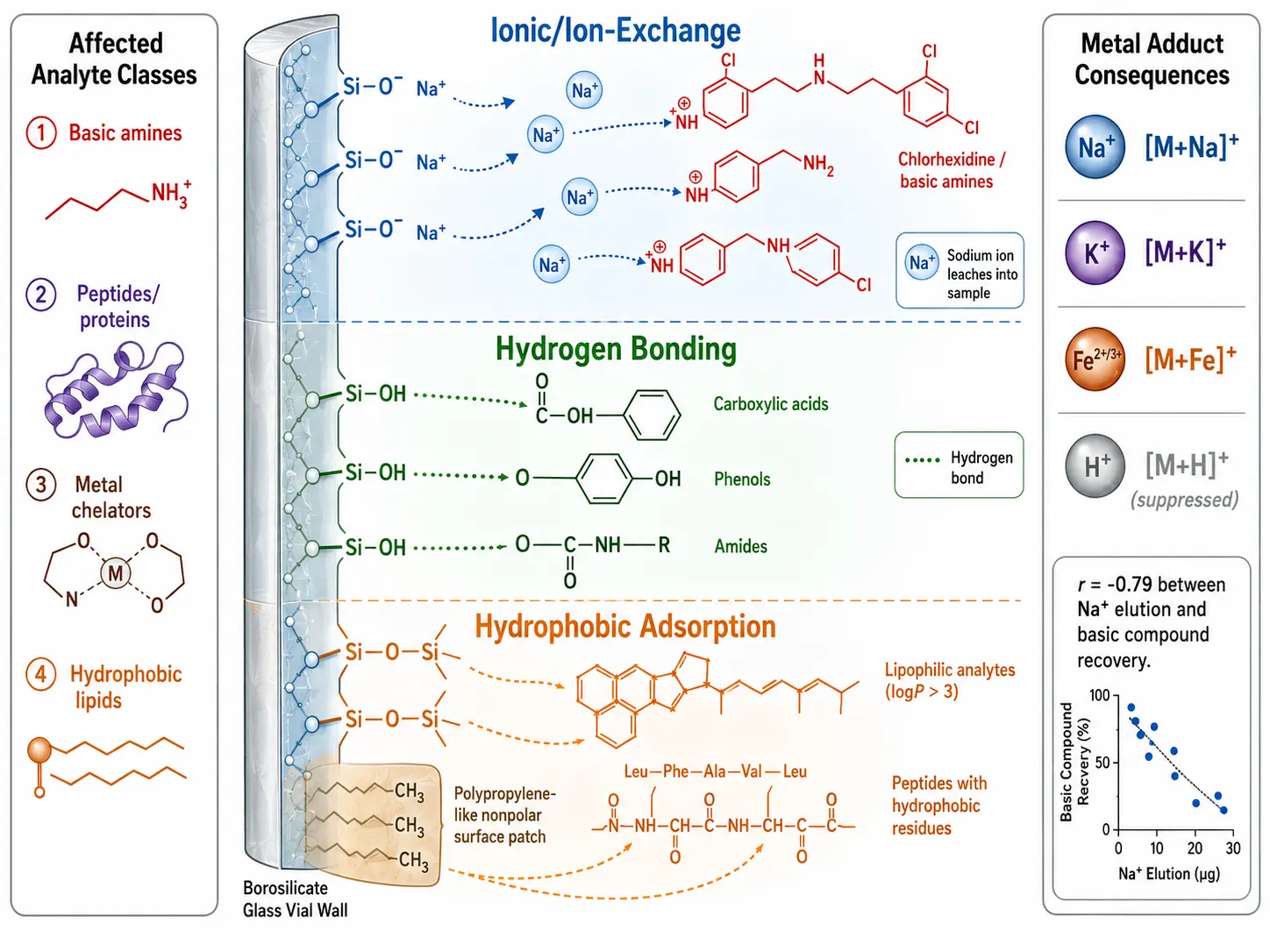 Figure 1: Mechanisms of Analyte Adsorption to Autosampler Vial Glass Surfaces
Mechanisms of Analyte Adsorption to Autosampler Vial Surfaces
Borosilicate glass — the standard material for autosampler vials — is not an inert container. Its surface chemistry actively interacts with dissolved analytes through three distinct mechanisms, each affecting different analyte classes with different kinetics. Understanding which mechanism is at play for a given analyte is the first step toward selecting an effective mitigation strategy.
Ion-Exchange Adsorption: The Sodium Connection
The dominant adsorption mechanism for basic compounds in glass vials is ion-exchange at deprotonated silanol groups. Borosilicate glass contains metal oxides — primarily sodium, but also potassium, calcium, aluminum, and iron — added during manufacturing to improve formability and chemical resistance. At the glass surface, these metals form metal silanolate groups (≡Si–O⁻ M⁺). When an aqueous or partially aqueous sample is introduced, the metal cations leach into solution, leaving behind negatively charged ≡Si–O⁻ sites. Protonated basic analytes — which carry a positive charge at the typical analytical pH of 2.5–4.0 — bind electrostatically to these sites, effectively extracting the analyte from solution onto the vial wall.
The quantitative relationship between sodium leaching and analyte recovery has been directly demonstrated. Osaka (2024) reported a correlation coefficient of r = −0.79 between sodium elution from 16 commercial vial products and chlorhexidine recovery after 72 hours of storage at 40°C. Vials that leached the most sodium showed the poorest recovery of this basic compound, with some products recovering less than 40% of the initial chlorhexidine concentration. Polypropylene vials — which lack silanol groups entirely — served as the inert control and maintained near-complete recovery. The same study demonstrated that sodium elution also promotes metal adduct formation in the electrospray ionization source: [M+Na]⁺, [M+K]⁺, and [M+Fe]⁺ adducts reduce sensitivity at the target [M+H]⁺ m/z channel, compounding the concentration loss from adsorption with an ionization efficiency loss.
Hydrogen Bonding to Neutral Silanols
Not all surface silanol groups are ionized. At the typical LC-MS injection solvent pH (2.5–4.0, corresponding to 0.1% formic acid in water-acetonitrile mixtures), a substantial fraction of silanol groups remain in the neutral ≡Si–OH form. These neutral silanols act as hydrogen bond donors and acceptors, interacting with polar functional groups on the analyte — carboxylic acids, phenols, amides, sulfonamides, and alcohols — through H-bond networks. Unlike ion-exchange, which is a high-affinity electrostatic interaction, hydrogen bonding is lower in energy per interaction. However, the high density of surface silanol groups (typically 4–5 Si–OH groups per nm² on fully hydroxylated glass) means that multi-point attachment can produce substantial cumulative binding, particularly for analytes with multiple hydrogen-bonding functional groups.
Hydrophobic Adsorption
Siloxane bridges (≡Si–O–Si≡) on the glass surface and the polymer backbone of polypropylene vials present hydrophobic patches that interact with lipophilic regions of analyte molecules. This mechanism predominates for analytes with logP > 3 and for peptides containing hydrophobic amino acid residues (leucine, isoleucine, valine, phenylalanine, tryptophan). Shimadzu's peptide adsorption studies using trypsin-digested myoglobin (~1.9 pmol/mL) revealed a polarity-dependent partitioning: highly polar peptides (retention time ~7–8 min on C18) preferentially adsorbed to glass vials via ion-exchange, while highly hydrophobic peptides (retention time ~12–16 min) preferentially adsorbed to polypropylene vials via hydrophobic interactions. The TORAST-H Bio vial, with its passivated polypropylene surface, minimized adsorption across both peptide classes simultaneously.
Analyte Classes at Highest Risk
The three mechanisms do not operate independently — most analytes interact with vial surfaces through some combination of all three, with one mechanism typically dominating based on the analyte's physicochemical properties. Basic amines and nitrogen-containing heterocycles (pKa > 7, positively charged at analytical pH) are primarily vulnerable to ion-exchange. Peptides and therapeutic proteins, with their combination of charged side chains, polar backbone amides, and hydrophobic residues, are vulnerable to all three mechanisms simultaneously — which is why 50–80% peptide loss to standard containers is commonly observed. Metal-chelating analytes (tetracyclines, fluoroquinolones, catechol-containing compounds) bind to surface metal ions through coordination chemistry, a mechanism distinct from but additive to the three primary pathways. Even neutral, moderately polar compounds can show measurable adsorption over extended storage (>24 hours) if the vial surface area-to-volume ratio is high, as in low-volume inserts where 100 µL of sample contacts a disproportionately large glass surface.
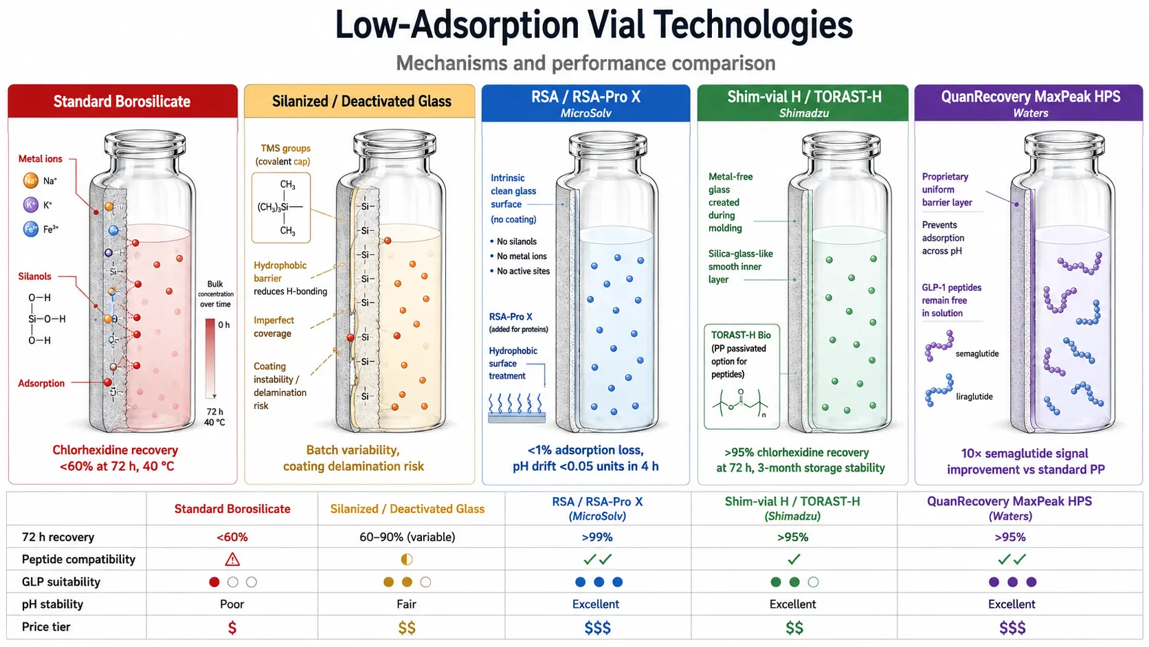 Figure 2: Commercial Low-Adsorption Vial Technologies — Mechanism Comparison
Commercial Low-Adsorption Vial Technologies: A Comparative Analysis
The autosampler vial market now offers several technology categories that address the adsorption problem through fundamentally different mechanisms — metal removal, covalent surface passivation, barrier coatings, and polymer-based alternatives. Selecting the right category and product for a specific analyte class and regulatory context requires understanding how each technology works at the molecular level.
Silanized (Deactivated) Glass Vials
Silanization is the oldest and most widely available surface deactivation method. The process reacts surface silanol groups with organosilane reagents — typically hexamethyldisilazane (HMDS), trimethylchlorosilane (TMSCl), or dimethyldichlorosilane (DMDCS) — to form covalent ≡Si–O–Si(CH₃)₃ bonds. The resulting trimethylsilyl surface is hydrophobic and chemically inert, blocking both ion-exchange and hydrogen bonding. Gas-phase silanization produces more uniform coverage than liquid-phase methods and is preferred for LC-MS vials.
However, silanized vials carry several limitations. Coverage is never complete — residual unreacted silanol groups remain, and incomplete coverage can produce vial-to-vial variability in adsorption behavior. The organosilane coating is susceptible to hydrolysis in aqueous solutions, with measurable degradation after approximately 48 hours. The silanization process can leave trace chloride residues (from chlorosilane reagents) that appear in LC-MS blanks. And the hydrophobic surface created by trimethylsilyl capping can, counterproductively, increase hydrophobic adsorption of lipophilic analytes even as it solves the ion-exchange problem.
RSA and RSA-Pro X (MicroSolv/MTC)
RSA (Reduced Surface Activity) vials take a fundamentally different approach: rather than coating the glass surface, the manufacturing process itself eliminates surface silanols and metal ions. The proprietary glass production process removes sodium, calcium, aluminum, and boron at the molding stage, producing a surface with 200–300× lower metal content than standard "certified" borosilicate vials. Because no coating is applied, there is nothing to delaminate, degrade, or vary in coverage — an important advantage for regulated bioanalytical workflows where vial-to-vial consistency is expected across thousands of injections.
Published performance data document less than 1% adsorption loss compared to up to 55% loss with untreated borosilicate glass, and pH drift of less than 0.05 units after 4 hours compared to up to 1.5 units for competitor vials. The RSA-Pro X variant adds a hydrolytically stable hydrophobic surface treatment specifically designed for proteins and peptides, remaining intact in 100% aqueous conditions and compatible with autoclaving and cryogenic storage to −80°C. RSA-Pro X vials are available in Max Recovery (center-draining, ~8 µL residual volume) and MRQ (~2 µL residual volume) geometries for limited-sample applications.
Shim-vial H and TORAST-H (Shimadzu)
Shimadzu's vial portfolio addresses adsorption through metal removal at the glass surface, similar to RSA in concept but executed through a multi-step cleaning and surface treatment process applied to USP Type 1 borosilicate glass during molding. The treatment removes alkali and transition metals from the surface layer, creating an effectively silica-glass-like inner surface that minimizes metal silanolate formation.
Osaka (2024) published a head-to-head comparison of 16 commercial vial products. Shim-vial H consistently showed the lowest sodium elution and the highest recovery of chlorhexidine — a basic compound chosen as a worst-case test probe — maintaining greater than 95% recovery after 72 hours at 40°C. The same study demonstrated that meloxicam (a weakly acidic NSAID) showed the highest proportion of [M+H]⁺ and the lowest [M+Na]⁺ adduct formation in Shim-vial H compared to all other glass vials tested, directly linking metal removal to improved MS sensitivity.
The TORAST-H product line extends this concept in two directions. TORAST-H Glass vials are validated for 3-month storage stability, with published data showing superior recovery maintenance compared to both standard polypropylene and competing glass vials over this period — making them the best-documented option for studies requiring long-term extract archiving. TORAST-H Bio vials are surface-passivated polypropylene designed specifically for peptide and protein analysis, where the hydrophobic polypropylene surface (which can adsorb lipophilic peptides) is rendered inert by the passivation treatment. Both products ship with lot-specific quality certificates documenting low-adsorption performance.
QuanRecovery with MaxPeak HPS (Waters)
Waters' QuanRecovery vials and plates with MaxPeak High Performance Surfaces (HPS) use a proprietary barrier coating technology distinct from both metal removal and silanization. The barrier layer prevents non-specific binding of proteins and peptides to the container surface without introducing the hydrophobic penalty of traditional silanization.
A March 2025 Waters application note (Trudeau and Lauber, 720008717) demonstrated the technology's performance with GLP-1 receptor agonist peptides — semaglutide, liraglutide, exenatide, and tirzepatide — which are large (3,700–4,800 Da), hydrophobic molecules with fatty acid conjugates that make them highly prone to surface adsorption. Compared to standard polypropylene plates, QuanRecovery MaxPeak HPS plates produced approximately 10× higher signal for semaglutide and approximately 8× higher signal for exenatide at 1 ng/mL concentration. These recovery improvements directly translate to lower achievable LLOQs, making the technology particularly relevant for peptide pharmacokinetic studies where sensitivity is the limiting factor.
Selection Framework
The appropriate vial technology depends on the analyte class, required storage duration, and regulatory context. For routine small-molecule LC-MS bioanalysis with acidic or neutral analytes, Shim-vial H or RSA vials provide cost-effective adsorption prevention with minimal lot-to-lot variability. For basic small molecules and metal-sensitive analytes, Shim-vial H is the best-documented option based on published head-to-head sodium elution and recovery data. For therapeutic peptides and proteins, QuanRecovery MaxPeak HPS offers the strongest published peptide recovery data, with RSA-Pro X and TORAST-H Bio as validated alternatives. For studies requiring multi-month extract archiving and re-analysis, TORAST-H Glass vials uniquely provide 3-month stability documentation. For GLP-regulated bioanalysis, all four technologies — RSA, Shim-vial H, TORAST-H, and QuanRecovery — provide the lot-specific quality certificates and traceability that regulatory submissions expect.
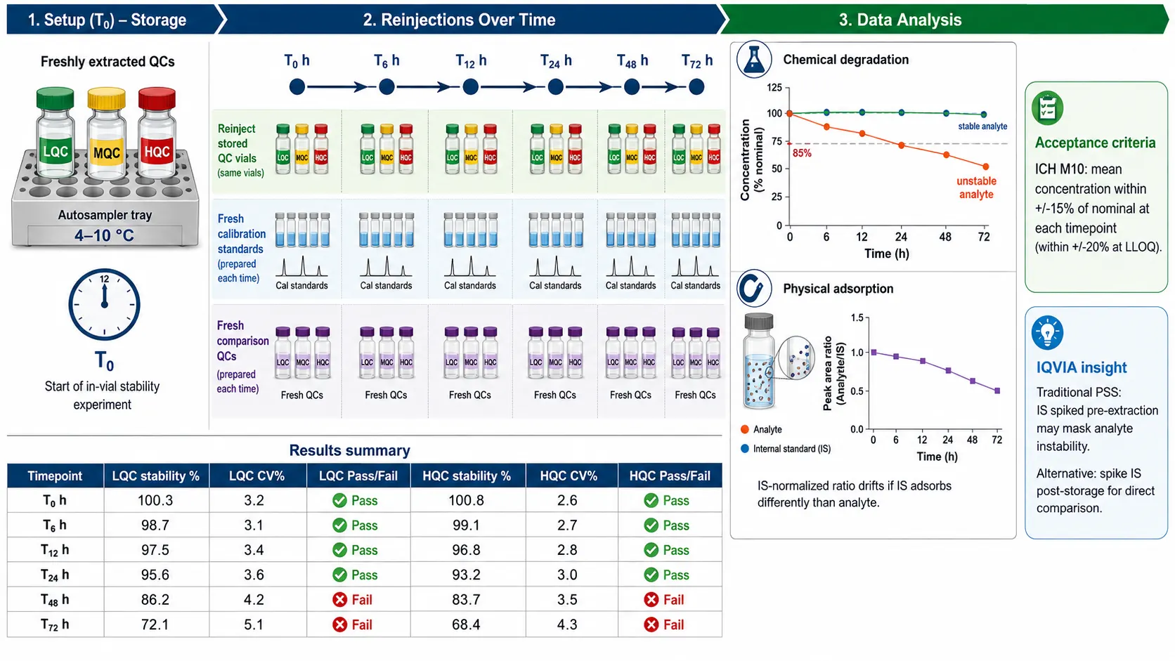 Figure 3: In-Vial (Autosampler) Stability Study Design — ICH M10-Compliant Protocol
In-Vial Stability Study Design and Acceptance Criteria
The in-vial stability experiment — also called autosampler stability, processed sample stability, or post-extraction stability — is a mandatory component of ICH M10 bioanalytical method validation. Its purpose is to demonstrate that processed samples stored under autosampler conditions (typically 4–10°C) remain quantitatively stable for the maximum duration of an analytical run, including time for potential re-injection after instrument downtime.
ICH M10 Requirements
The ICH M10 guideline on bioanalytical method validation, adopted in May 2022 and implemented January 2023, harmonizes FDA and EMA regional guidances and explicitly requires stability demonstration for the analyte in processed samples. The guideline expects that stability be evaluated using low and high QC concentration levels, with the tested storage duration bracketing the maximum anticipated batch runtime. The acceptance criterion is consistent with general ICH M10 accuracy/precision requirements: the mean back-calculated concentration at each stability time point should be within ±15% of the nominal concentration (or within ±20% at the LLOQ). For a 96-well plate batch with 10-minute LC-MS run times per sample, the maximum queue duration is approximately 16 hours, so 24-hour stability documentation is the practical minimum, with 48–72 hours recommended to accommodate instrument downtime and re-injection scenarios.
Experimental Design: The Traditional Approach and Its Limitations
The conventional post-extraction stability experiment proceeds as follows: QC samples at low and high concentrations are extracted, reconstituted, and transferred to autosampler vials. One set is injected immediately (T₀) against a freshly prepared calibration curve. The remaining vials are stored in the autosampler at the setpoint temperature (commonly 4–10°C) and re-injected at pre-defined intervals — typically 6, 12, 24, and 48 hours — against fresh calibration curves prepared at each time point. Stability is calculated as the ratio of the mean back-calculated concentration at each time point to the mean concentration of freshly prepared comparison QCs.
However, as IQVIA Laboratories highlighted in a 2023 technical blog, this traditional design carries a structural flaw. When the internal standard is spiked into the sample before extraction, any IS stability issues during post-extraction storage are indistinguishable from analyte stability issues. A stable isotope-labeled IS (SIL-IS) that degrades or adsorbs at the same rate as the analyte will mask true instability by maintaining a constant analyte/IS peak area ratio. Conversely, if the IS adsorbs to the vial surface differently than the analyte, the IS-normalized response will drift in ways that the calibration curve cannot correct, potentially producing a false stability failure or, worse, a false pass. An alternative approach — spiking the IS into the processed extract after the storage period rather than before — decouples IS stability from analyte stability and provides a direct measurement of analyte recovery, though it sacrifices the quantitative normalization that IS provides during the extraction step.
Re-Injection Reproducibility
ICH M10 also requires demonstration of re-injection reproducibility — the ability to re-process and re-inject an entire batch of extracted samples after a realistic delay (typically 24–72 hours) and obtain results within pre-specified acceptance limits of the original injection. This experiment validates both the instrument's analytical reproducibility and the processed sample's chemical/physical stability over the re-injection window, and is considered by many bioanalysts to be the more practically informative of the two post-extraction stability experiments. Published validation studies routinely demonstrate re-injection reproducibility with CV values below 3% at both low and high QC concentrations over 48–77 hour windows when appropriate vial technology and solvent conditions are used.
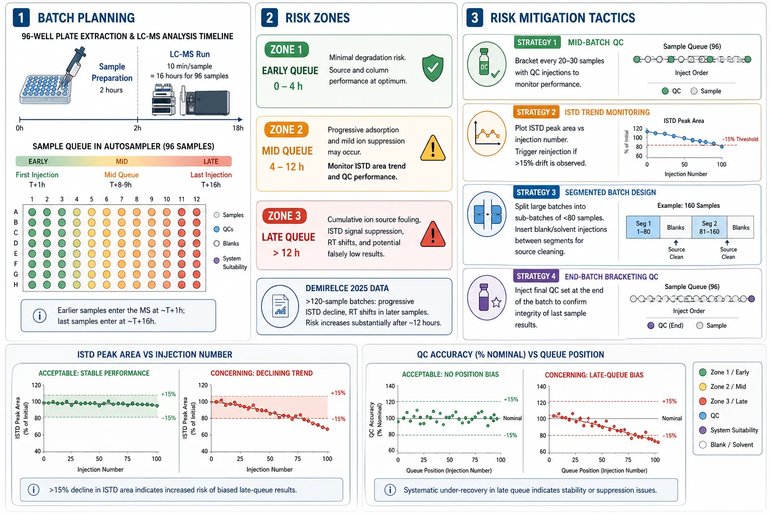 Figure 4: High-Throughput Queuing and Large-Batch Stability Risk Management
High-Throughput Queuing Strategies for Large Analytical Batches
As bioanalytical laboratories push throughput to meet the demands of drug discovery programs — where a single PK study may generate 96 to 384 samples requiring results within 24–48 hours — the practical management of autosampler queue time becomes a critical variable. A 96-well plate extracted and queued for LC-MS analysis at 10 minutes per sample represents a 16-hour analytical run. The first sample sits in the autosampler for approximately 1 hour after extraction; the last sample sits for 16 hours.
The Large-Batch Signal Drift Phenomenon
Demirelce (2025) documented a systematic phenomenon in clinical LC-MS/MS analysis: while small batches (20–30 samples) showed stable internal standard peak areas and consistent retention times throughout the run, large batches (≥120 samples) exhibited progressive internal standard signal decline, slight retention time shifts, and — critically — falsely low measured concentrations in the later portions of the batch. The attributed mechanisms include cumulative deposition of endogenous residues on the ion source and early column section, and subtle temperature variations in the autosampler tray during prolonged operation affecting analyte/solvent distribution and evaporation rates. These effects are distinct from, and additive to, the vial adsorption mechanisms discussed above.
Risk Mitigation Through Batch Design
Four operational strategies can reduce the risk of queue-time-dependent data degradation without sacrificing throughput. First, mid-batch QC samples — bracketed quality control injections placed every 20–30 samples — provide real-time monitoring of batch integrity. If the QC accuracy at position 60 falls below the 85–115% acceptance window while QCs at position 20 remain within limits, the operator has early warning of a progressive problem and can halt the run before generating invalid data. Second, internal standard peak area trend monitoring — plotting the IS peak area against injection number for every sample in the batch — provides a continuous readout of ion source and system health. A systematic downward trend exceeding 15% from the first to the last injection is grounds for investigation, even if individual QC values remain within acceptance limits. Third, segmented batch design divides large batches into sub-batches of 60–80 samples, with blank and solvent injections interspersed between sub-batches to allow brief ion source recovery. Fourth, end-batch bracketing QC — a final set of QC samples injected after the last study sample — provides direct evidence that analytical performance was maintained through the final injection.
Autosampler Temperature Management
The autosampler temperature setpoint — typically 4°C or 10°C — represents a compromise between stability (lower is better) and chromatography (very cold samples can cause peak broadening due to solvent viscosity effects and thermal mismatch at the column head). For most small-molecule LC-MS methods, 10°C provides adequate stability for 24–48 hours without chromatographic penalty. For thermally labile analytes or long queue durations (>48 hours), 4°C is preferred, with acceptance that a brief column re-equilibration period after injection may be needed. Regardless of the chosen setpoint, the autosampler temperature must be verified to remain within ±2°C of the setpoint throughout the entire batch duration, and the in-vial stability experiment must be conducted at the same temperature used for study sample analysis.
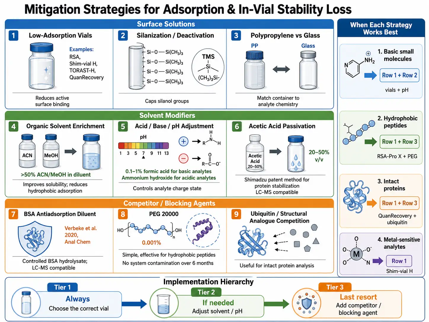 Figure 5: Adsorption Mitigation Strategy Toolbox for LC-MS Bioanalysis
Adsorption Mitigation Strategies: Vials, Solvents, and Competitive Agents
When an analyte shows evidence of post-extraction adsorption — declining peak areas over time in the autosampler, poor replicate precision at low concentrations, or concentration-dependent recovery — a tiered mitigation approach is recommended, starting with the simplest and most robust interventions and escalating only as needed.
Tier 1: Vial Selection (Always First)
The single most effective intervention for most adsorption problems is switching to an appropriate low-adsorption vial. The mechanism of adsorption should guide the choice: ion-exchange of basic compounds → Shim-vial H or RSA (both eliminate surface metal silanolates); hydrophobic adsorption of lipophilic analytes → silanized glass or TORAST-H (hydrophobic surface minimizes aqueous-phase hydrophobic interactions); peptide/protein multi-mechanism adsorption → QuanRecovery MaxPeak HPS, RSA-Pro X, or TORAST-H Bio. Our bioanalysis of challenging compounds and complex matrices services incorporate systematic vial screening as part of method development for adsorption-prone analytes.
Tier 2: Solvent Modification (If Vial Change Is Insufficient)
The reconstitution solvent composition directly affects both analyte solubility and surface interaction energetics. Three solvent modification strategies have broad applicability. Organic solvent enrichment — maintaining at least 50% acetonitrile or methanol in the final injection solvent — improves solubility of lipophilic analytes and reduces hydrophobic adsorption by competing for hydrophobic surface sites. For HPLC methods where high organic content in the injection solvent causes peak broadening (solvent strength mismatch with the initial gradient conditions), a 50 µL injection volume or smaller mitigates this effect on standard 2.1 mm ID columns. pH control — ensuring the analyte is in its most soluble ionization state — can reduce electrostatic adsorption: protonated basic analytes bind to silanolates, so raising the pH above the analyte's pKa (within the constraints of chromatographic performance and analyte stability) reduces the fraction of positively charged analyte available for ion-exchange. Acetic acid at 20–50% (v/v) in the sample diluent has been shown in a Shimadzu patent (US 2021/0302286 A1) to prevent protein adsorption to glass and plastic surfaces without the LC-MS interference that conventional surfactants produce.
Tier 3: Competitive Adsorption Agents (For Stubborn Cases)
When vial and solvent interventions are insufficient — most commonly for therapeutic peptides, ultra-trace analytes (sub-ng/mL), or methods that constrain the injection solvent composition — competitive adsorption agents can be added to the sample to saturate surface binding sites and keep the analyte in solution. Three well-characterized options have been published.
The BSA-based antiadsorption diluent (Verbeke et al., 2020, Analytical Chemistry) uses controlled hydrolysis and precipitation of bovine serum albumin to produce an LC-MS-compatible peptide mixture that competitively occupies vial surface sites. BSA is dissolved at 10 mg/mL in water with 0.1% formic acid, diluted to 2.5 mg/mL with acidified acetonitrile, heated at 95°C for 5 minutes, cooled on ice, and centrifuged. The supernatant — containing BSA hydrolysis products but no intact protein that would foul the LC-MS system — serves as the antiadsorption diluent and is fully compatible with formic acid and acetonitrile addition for method development flexibility.
Polyethylene glycol (PEG 20000) at 0.001% (w/v) is a simpler alternative that does not require preparation. Research from the Zdráhal group (Masaryk University) demonstrated that 0.001% PEG 20000 in the injection solution increased peptide identifications by 34–64% compared to formic acid-only solutions in proteomics workflows, with no adverse effects on the LC-MS system over six months of continuous use. PEG is particularly effective at preserving longer, more hydrophobic peptides (retention time > 25 min on C18) that are preferentially lost to hydrophobic vial surfaces.
The comprehensive review by Maes et al. (2014, Journal of Chromatography A) catalogs additional adsorption competitors — including rat plasma (0.05%), Fmoc-arginine, poly(ethylenimine), glucagon, and structural analogue peptides — with practical guidance on selecting an appropriate competitor based on analyte properties and analytical constraints. The key principle across all competitive approaches is that the competitor must be present at sufficient concentration to saturate surface binding sites (typically orders of magnitude higher than the analyte concentration) while remaining chromatographically resolved from the analyte and not causing ion suppression at the analyte's retention time.
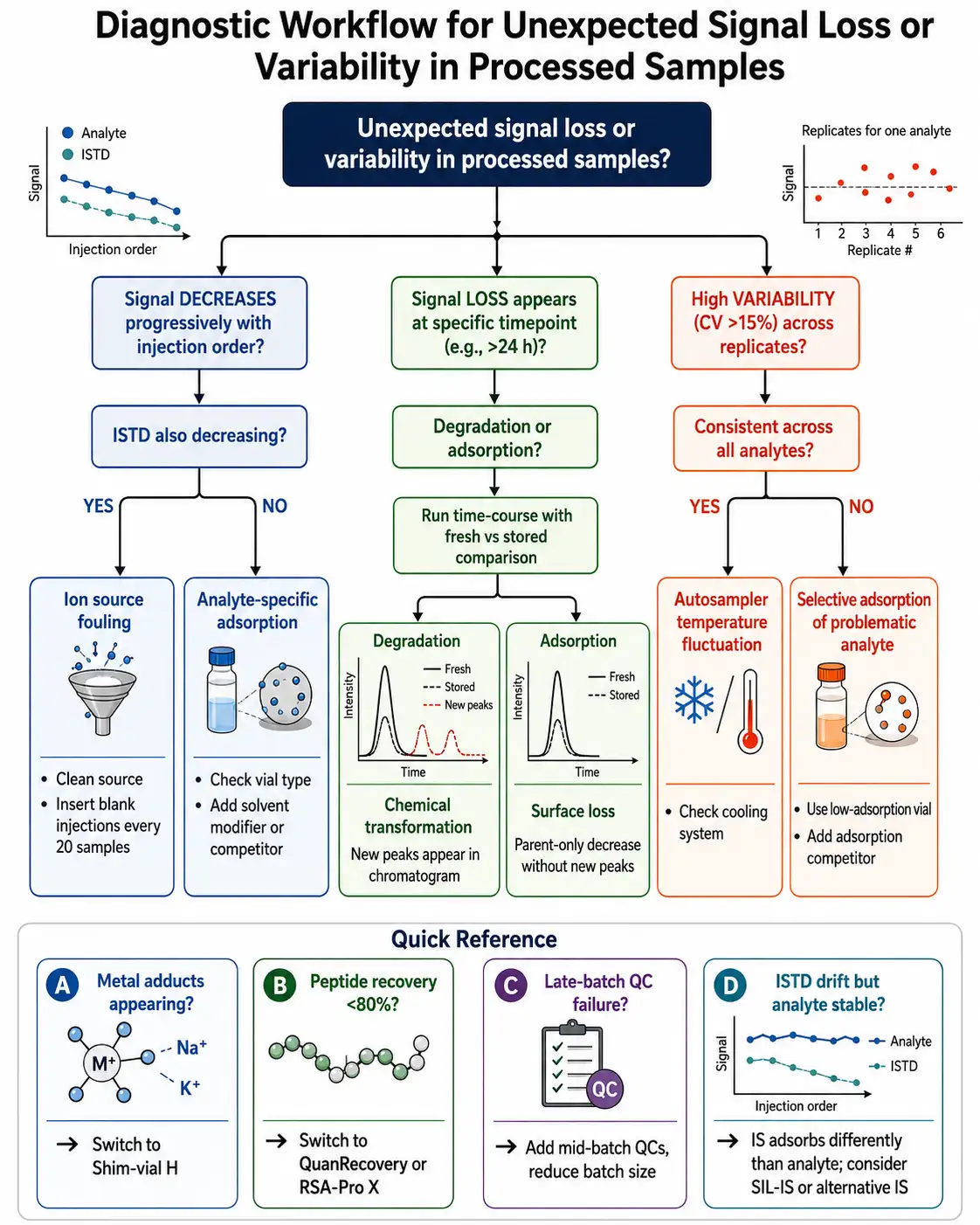 Figure 6: Post-Extraction Sample Integrity Troubleshooting Decision Tree
Troubleshooting Post-Extraction Recovery Issues
When processed sample recovery problems arise — whether during method development, validation, or routine study sample analysis — a systematic diagnostic approach identifies the root cause more efficiently than trial-and-error substitution of vials and solvents.
Symptom 1: Progressive Signal Decline with Injection Order
If both analyte and internal standard peak areas decline progressively over the course of a batch, the cause is likely ion source fouling or column contamination rather than vial adsorption. In this scenario, the analyte/IS peak area ratio remains stable because both decline together. Solution: clean the ion source, insert blank/solvent injections every 20–30 samples to flush the column and source, or reduce the batch size. If the analyte peak area declines but the IS peak area remains stable, selective vial adsorption of the analyte is the more likely cause — the IS may be less surface-active due to different physicochemical properties. Switching to a SIL-IS that matches the analyte's adsorption behavior is the preferred solution when feasible.
Symptom 2: Time-Dependent Loss at Specific Storage Duration
If recovery is acceptable at early time points (0–12 hours) but drops below the 85% threshold at later time points (24–72 hours), the rate of loss must be characterized to distinguish degradation from adsorption. Degradation typically produces new chromatographic peaks (degradation products) and is temperature-dependent (faster at higher autosampler temperatures). Adsorption produces a decrease in the parent peak without new peaks appearing and may reach a plateau when surface binding sites become saturated — recovery stabilizes at some sub-100% level rather than continuing to decline indefinitely. Running a parallel experiment in polypropylene vials (which lack silanol groups but may still show hydrophobic adsorption) versus low-adsorption glass vials can quickly distinguish the dominant mechanism.
Symptom 3: High Variability Across Replicates at Low Concentration
Poor precision (CV > 15%) at the LLOQ level that is not observed at higher QC concentrations, combined with lower-than-expected recovery, is the classic signature of surface adsorption. The surface area-to-analyte ratio is most unfavorable at low concentrations, where a larger fraction of the total analyte mass can be removed by a fixed number of surface binding sites. Switching to a low-adsorption vial with smaller internal surface area (a low-volume insert rather than a full-size vial, or a center-draining Max Recovery geometry) often resolves this problem by simultaneously reducing both the total binding site count and the surface area-to-volume ratio.
Symptom 4: Metal Adduct Peaks Competing with [M+H]⁺
If the mass spectrum shows prominent [M+Na]⁺, [M+K]⁺, or [M+Fe]⁺ peaks alongside — or instead of — the intended [M+H]⁺ peak, the vial is contributing metal ions to the sample solution. The solution is to switch to a vial technology that removes surface metals during manufacturing (Shim-vial H or RSA) rather than silanized vials, which cap silanols but do not address the underlying metal content of the glass. In the short term, adding 0.1% formic acid to the injection solvent can suppress sodium adduct formation by providing abundant protons to compete with sodium during electrospray ionization, though this addresses the symptom rather than the source of sodium.
For method-specific troubleshooting of challenging analytes, our bioanalytical method development and validation services and sample preparation method development services provide systematic investigation of analyte-specific adsorption behavior and tailored mitigation strategies. For stability studies spanning the full range of storage conditions, our short-term and long-term stability studies in bioanalysis services cover bench-top, freeze-thaw, autosampler, and long-term frozen storage evaluation under ICH M10-compliant protocols.
Frequently Asked Questions About Post-Extraction Sample Integrity
How long can processed samples safely remain in the autosampler before re-injection?
ICH M10 requires that the validated post-extraction stability period covers the maximum anticipated batch runtime, including potential re-injection delays. For a typical 96-well plate with 10-minute run times (~16 hours), 24-hour stability documentation is the practical minimum; 48–72 hours is recommended for labs that may need to re-queue batches after instrument downtime. The actual stability window depends on the analyte, injection solvent, vial type, and autosampler temperature. Published validations routinely achieve 48–77 hour autosampler stability with CV below 3% when appropriate vial technology and solvent conditions are used.
What is the difference between silanized vials and RSA/Shim-vial H type vials?
Silanized vials apply a reactive organosilane coating (typically trimethylsilyl groups) to cap surface silanol groups after the vial is formed. The coating is covalent but can be incomplete, hydrolytically unstable in aqueous solutions beyond ~48 hours, and may introduce trace chloride contamination. RSA and Shim-vial H vials remove the metal ions that form silanolates at the glass surface during the manufacturing process itself — no coating is applied. This eliminates delamination risk, provides more consistent vial-to-vial performance, and addresses the root cause (metal content) rather than the symptom (exposed silanols).
Why do peptides and proteins require special vial considerations compared to small molecules?
Peptides and proteins are vulnerable to all three vial adsorption mechanisms simultaneously: charged side chains (Lys, Arg, His, Asp, Glu) participate in ion-exchange at silanolate sites; the peptide backbone amide bonds and polar side chains engage in hydrogen bonding with neutral silanols; and hydrophobic residues (Leu, Ile, Val, Phe, Trp) drive adsorption to siloxane and polypropylene surfaces. This multi-mechanism vulnerability explains the 50–80% peptide losses commonly observed with standard containers. Dedicated peptide-grade vials — QuanRecovery MaxPeak HPS, RSA-Pro X, or TORAST-H Bio — address all three mechanisms simultaneously through barrier coatings or combined metal removal and surface passivation.
Can I add BSA or PEG to my samples without affecting LC-MS performance?
Native (intact) BSA cannot be used directly — it will foul the LC column and ion source. The Verbeke et al. (2020) BSA antiadsorption diluent uses controlled hydrolysis (95°C, 5 min in acidified conditions) followed by centrifugation to remove intact protein, producing a peptide-based competitor that is LC-MS compatible. PEG 20000 at 0.001% (w/v) has been demonstrated to produce no adverse system effects over six months of continuous use and is the simpler option for most workflows. Both approaches should be validated for the specific analyte and method: spike the competitor into QC samples, compare recovery and precision against competitor-free samples, and confirm that no new chromatographic peaks or ion suppression zones appear at the analyte's retention time.
How do I know if my internal standard is masking post-extraction instability?
If both the analyte and IS are declining at the same rate during post-extraction storage, the analyte/IS peak area ratio stays constant and the stability result appears acceptable — even though the absolute analyte signal is dropping. This is most likely with SIL-IS, which matches the analyte's chemical behavior. To test for this masking effect: (1) compare the absolute analyte peak area (not the ratio) at T0 versus the last stability time point; (2) run a parallel experiment where IS is spiked after storage rather than before extraction, comparing the stored samples against a calibration curve prepared with post-storage-spiked IS. If the absolute analyte peak area declines by more than 15% while the IS-normalized result stays within limits, the IS is masking real analyte instability and mitigation is needed even though the traditional acceptance criteria are met.
What is the most cost-effective vial strategy for a high-throughput discovery lab running 500+ samples per week?
For high-throughput discovery workflows, the cost-optimized strategy is to categorize analytes by adsorption risk and assign vial types accordingly. The majority of small-molecule drugs with moderate logD (0–3) and pKa below 8 show minimal adsorption in standard borosilicate vials over typical discovery batch durations (<12 hours) and do not require premium vials. Reserve Shim-vial H or RSA vials for the subset of compounds showing evidence of adsorption — basic amines (pKa > 8), metal chelators, and analytes with LLOQ below 1 ng/mL where even minor losses impact quantification. Reserve QuanRecovery or RSA-Pro X for peptide and protein therapeutics. This risk-based approach allocates premium vial cost to the roughly 15–25% of compounds that benefit from it, rather than applying a uniform premium-vial policy to all samples. Our LC-MS/MS single drug quantification services incorporate this tiered vial strategy to balance cost and data quality across discovery programs.
References
- ICH M10 Guideline: Bioanalytical Method Validation and Study Sample Analysis. International Council for Harmonisation of Technical Requirements for Pharmaceuticals for Human Use; adopted May 2022, implemented January 2023. https://www.ich.org/page/multidisciplinary-guidelines
- Maes K, Smolders I, Michotte Y, Van Eeckhaut A. Strategies to reduce aspecific adsorption of peptides and proteins in liquid chromatography-mass spectrometry based bioanalyses: An overview. Journal of Chromatography A. 2014;1358:1-13. DOI: 10.1016/j.chroma.2014.06.072
- Verbeke F, Bracke N, Debunne N, Wynendaele E, De Spiegeleer B. LC-MS Compatible Antiadsorption Diluent for Peptide Analysis. Analytical Chemistry. 2020;92(2):1712-1719. DOI: 10.1021/acs.analchem.9b01840
- Osaka Y. Improved Data Reliability with New Low-Adsorption Vials. LC to LC/MS no Chie. 2024;8:30-35. Shimadzu Corporation.
- Trudeau M, Lauber MA. Improved GLP-1 Receptor Agonist Peptide Recovery Using a QuanRecovery with MaxPeak High Performance Surfaces (HPS) Collection Plate. Waters Corporation Application Note 720008717; March 2025. Waters Application Note 720008717
- Demirelce O. Large batch size effect on signal stability and retention time consistency in LC-MS/MS analyses: A technical observation. The Injector. 2025;4(3):106-107. DOI: 10.5281/zenodo.19209276
- United States Pharmacopeia. USP General Chapter <660> Container-Glass. USP-NF. Rockville, MD: United States Pharmacopeial Convention.
- IQVIA Laboratories. Post Extracted Sample Stability (PSS) experiment and can't we do better? IQVIA Laboratories Blog; 2023. https://labs.iqvia.com/blog/post-extracted-sample-stability-experiment-cant-we-do-better
Related Services


