Reactive Metabolite Formation and Idiosyncratic Drug Reaction Mechanisms
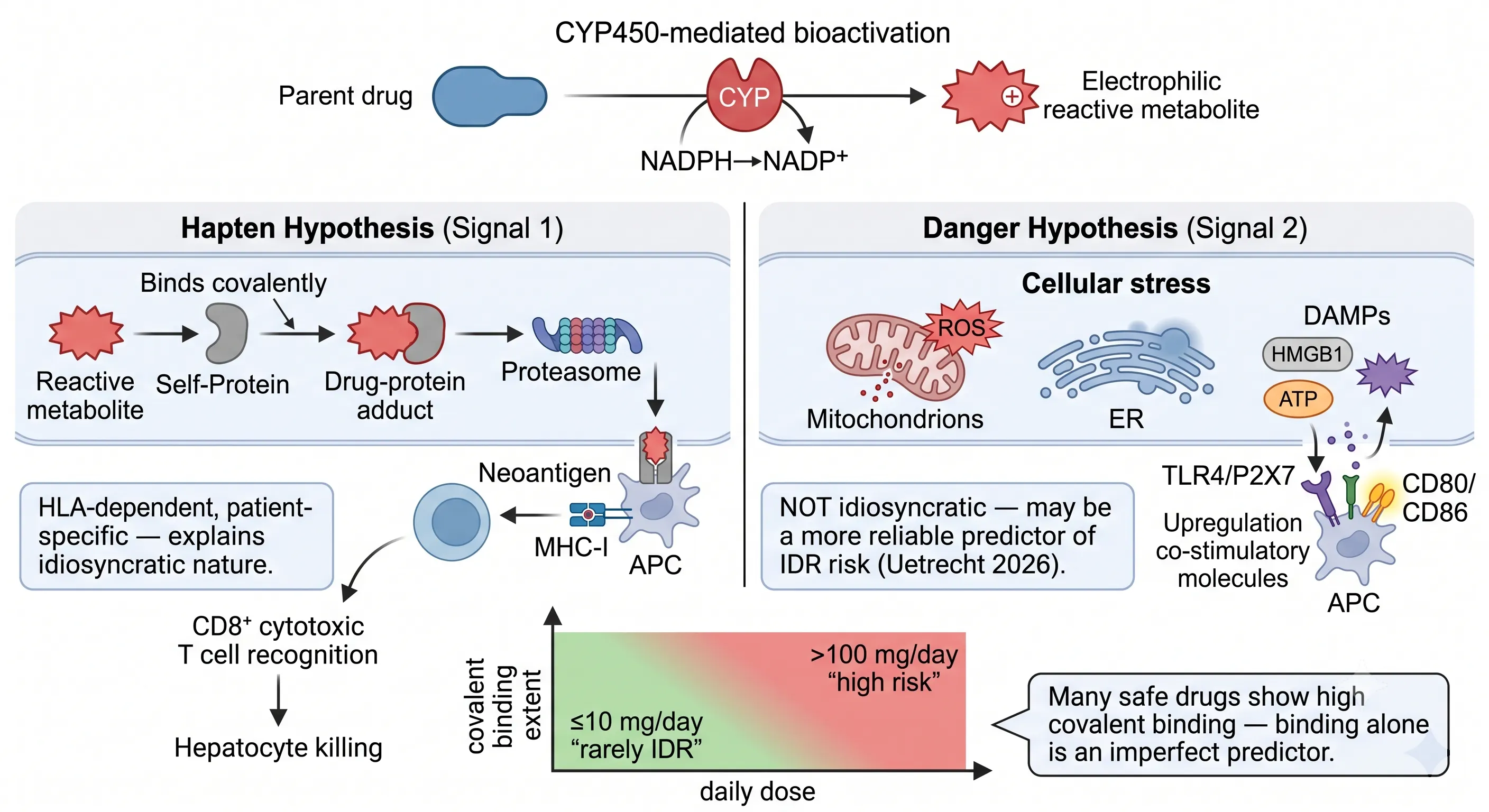 Figure 1: Reactive Metabolite Formation and Idiosyncratic Drug Reaction Mechanisms — Hapten vs Danger Hypothesis
A reactive metabolite is not a toxicologist's curiosity — it is the molecular initiating event for most idiosyncratic drug reactions (IDRs), and every drug discovery program that overlooks reactive metabolite screening is accepting a risk that no amount of preclinical safety pharmacology can mitigate. IDRs are the leading cause of post-marketing drug withdrawal and black-box warnings, and they are fundamentally unpredictable from the parent drug's pharmacology because they are mediated by chemically reactive metabolites, not the parent compound itself.
The formation cascade is well-characterized. The parent drug — typically a chemically inert, pharmacologically optimized molecule — enters hepatocytes and encounters CYP450 enzymes. CYP-mediated oxidation converts the drug into an electrophilic reactive metabolite: a chemical species bearing a partial or full positive charge, an unpaired electron, or an electron-deficient center that seeks nucleophilic attack. Under normal conditions, this electrophile is quenched by glutathione (GSH) and excreted harmlessly. But when the reactive metabolite escapes GSH conjugation — because it is formed at a rate exceeding GSH availability, or because its electrophilicity is directed toward harder nucleophiles than the soft thiol of GSH — it attacks cellular macromolecules: proteins, DNA, and membrane lipids.
The downstream consequences are explained by two complementary but mechanistically distinct hypotheses. The hapten hypothesis (Signal 1): the reactive metabolite covalently binds to a self-protein, forming a drug-protein adduct. This adduct is processed by the proteasome, presented as a neoantigen on MHC-I molecules by antigen-presenting cells (APCs), and recognized by CD8+ cytotoxic T cells, which initiate hepatocyte killing. This pathway is HLA-dependent and patient-specific — explaining why IDRs are rare, unpredictable, and not dose-proportional in the classical sense. Different individuals have different HLA alleles, and only certain HLA variants present a given drug-protein adduct in a conformation that T cells recognize. The danger hypothesis (Signal 2): the reactive metabolite causes direct cellular stress — mitochondrial damage with reactive oxygen species (ROS) release, endoplasmic reticulum stress, and the extrusion of damage-associated molecular patterns (DAMPs) including HMGB1 and ATP. These DAMPs bind Toll-like receptor 4 (TLR4) and P2X7 purinergic receptors on APCs, triggering upregulation of co-stimulatory molecules (CD80/CD86) that provide the essential "Signal 2" for full T cell activation. Without Signal 2, even a drug-protein neoantigen presented on MHC-I does not trigger an immune response — the T cell becomes anergic rather than activated.
A 2026 review by Uetrecht systematically examined the evidence for both hypotheses and reached a conclusion with direct implications for drug discovery: the danger signal (Signal 2) may be a more reliable predictor of IDR risk than covalent binding alone (Drug Metab Rev, 2026). The reasoning is that covalent binding is necessary but not sufficient — many safe, widely prescribed drugs exhibit substantial covalent binding — while cellular stress and DAMP release appear to be the rate-limiting step that determines whether immune tolerance is broken. This insight is reshaping the reactive metabolite risk assessment paradigm, shifting emphasis from "how much covalent binding" toward "what type of cellular stress does this reactive metabolite cause."
At Creative Proteomics DMPK, reactive metabolite screening goes beyond trap-and-detect — every positive trapping result is interpreted in the context of the hapten and danger pathways, the drug's structural alert profile, and the projected clinical dose to provide a holistic RM risk assessment that directly informs candidate selection decisions.
GSH Trapping Methodology — From Microsomal Incubation to HRMS Detection
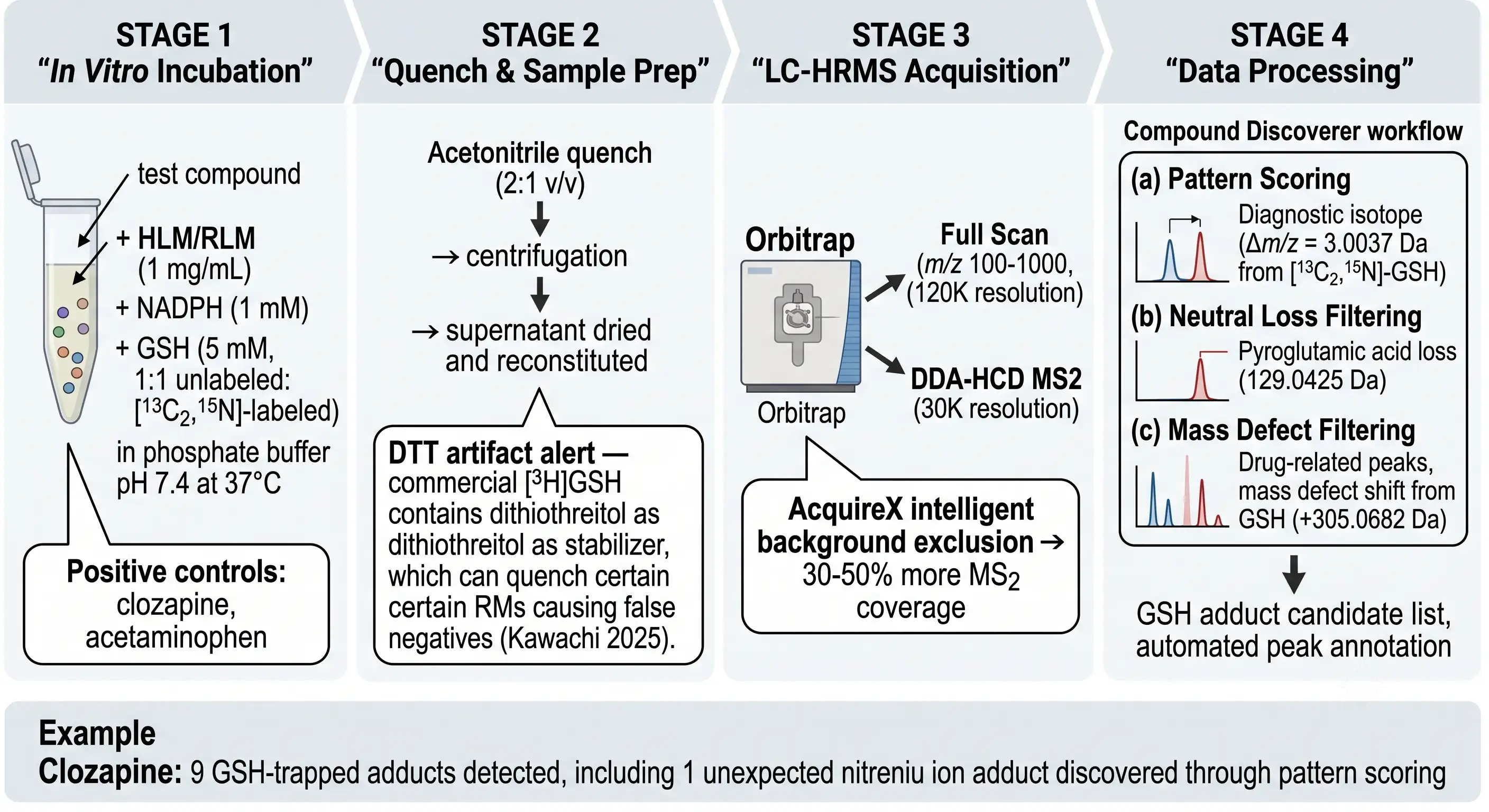 Figure 2: GSH Trapping Methodology — From Microsomal Incubation to HRMS Detection
GSH trapping is the workhorse reactive metabolite detection assay — it is the first-line screen applied to every lead compound in a well-designed DMPK cascade. The principle is straightforward: GSH (γ-glutamyl-cysteinyl-glycine) is the cell's endogenous electrophile scavenger, present at millimolar concentrations in hepatocytes. When incubated with drug-metabolizing enzymes at supraphysiological GSH concentrations (5 mM), any electrophilic reactive metabolite generated is trapped as a stable GSH adduct that can be detected, identified, and semi-quantified by LC-HRMS. If the assay is clean, the electrophile burden is low; if GSH adducts are detected, the structural alert and the adduct-forming mechanism must be understood before proceeding.
The standard GSH trapping incubation uses human liver microsomes (HLM, 1 mg/mL) or rat liver microsomes (RLM) with NADPH (1 mM) as the CYP cofactor and GSH (5 mM) as the trapping agent in phosphate buffer (pH 7.4) at 37°C. The critical methodological evolution since 2021 has been the transition from [35S]GSH to [3H]GSH as the radiolabeled trapping agent, due to the commercial discontinuation of 35S-GSH. This transition introduced an unanticipated artifact: commercial [3H]GSH contains dithiothreitol (DTT) as a stabilizer, and DTT is itself a nucleophile that can quench certain reactive metabolites before they react with GSH, producing false negatives. Kawachi et al. (2025) documented this DTT artifact and recommended that negative [3H]GSH trapping results be confirmed with an orthogonal method when the drug carries high-risk structural alerts (Drug Metab Pharmacokinet, 65:101504).
For discovery-stage screening where throughput and cost are prioritized over absolute quantitation, dansyl-GSH (dGSH) — a fluorescent GSH derivative — provides a non-radioactive alternative. dGSH (excitation 328-340 nm, emission 525-541 nm) forms fluorescent adducts with soft electrophiles, detectable by LC-fluorescence without the regulatory and infrastructure overhead of radiochemical work. The trade-off is that dGSH only traps soft electrophiles and has lower sensitivity than the radiometric method; it serves as a rapid first-pass screen, with positive hits escalated to [3H]GSH or HRMS-based methods for confirmation and structural characterization.
The LC-HRMS detection and data processing workflow has advanced substantially. Modern GSH trapping analysis uses three parallel detection algorithms operating on Orbitrap HRMS data (Full Scan m/z 100-1000 at 120K resolution, with DDA-HCD MS2 at 30K resolution): (1) pattern scoring detects the diagnostic isotope doublet from 1:1 unlabeled:[13C2,15N]-GSH (Δm/z = 3.0037 Da), flagging drug-related peaks; (2) neutral loss filtering identifies the characteristic loss of pyroglutamic acid (129.0425 Da) from GSH adducts in MS2 spectra; and (3) mass defect filtering identifies drug-related peaks with the specific mass defect shift from GSH conjugation (+305.0682 Da). Intelligent background exclusion (AcquireX on Thermo platforms) improves MS2 coverage by 30-50% by directing fragmentation time toward lower-abundance drug-related ions and away from background. The output is a GSH adduct candidate list with automated peak annotation, supported by Fragment Ion Search (FISh) scoring that matches experimental MS2 fragments against in silico predicted adduct fragment structures. The foundational principles of adduct structural characterization draw on the same HRMS fragmentation logic used in advanced drug metabolite identification, where MS2 spectral annotation and site-of-modification determination are covered in detail.
The Double Trapping Revolution — GSH + Cyanide for Comprehensive Electrophile Coverage
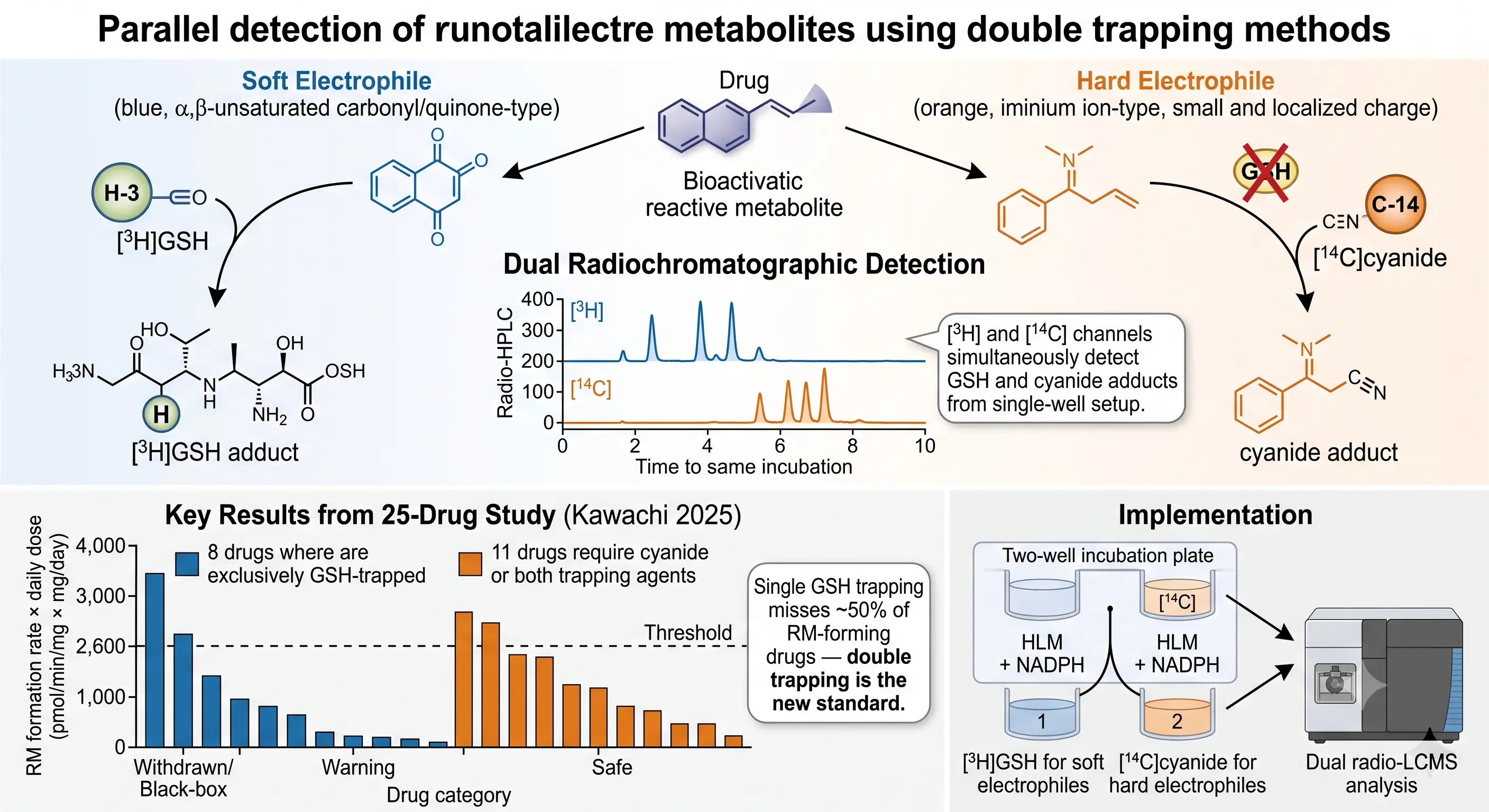 Figure 3: The Double Trapping Revolution — GSH + Cyanide for Comprehensive Electrophile Coverage
The single most important finding in reactive metabolite screening methodology in the past five years is that GSH trapping alone misses approximately 50% of RM-forming drugs. This is not a minor sensitivity issue — it means that a drug discovery program relying exclusively on GSH trapping is accepting a coin-flip probability of obtaining a false-negative RM assessment. The 2025 study by Kawachi et al. that established this figure tested 25 drugs across four risk categories (Withdrawn/Black-box, Warning, Safe) using both [3H]GSH and [14C]cyanide trapping, with dual-channel radio-HPLC detection, and found that 8 drugs were exclusively GSH-trapped (soft electrophiles only), while 11 drugs required cyanide trapping — either alone or in combination with GSH — for complete RM detection (Drug Metab Pharmacokinet, 65:101504).
The mechanistic explanation lies in the hard-soft acid-base (HSAB) principle. GSH is a soft nucleophile — its reactive thiol group (-SH) is highly polarizable and preferentially reacts with soft electrophiles: α,β-unsaturated carbonyls, quinones, quinone-imines, epoxides, and Michael acceptors. These "soft electrophiles" are the classical bioactivation products that medicinal chemists are trained to avoid. Cyanide is a hard nucleophile — its small, localized negative charge on carbon preferentially reacts with hard electrophiles: iminium ions (R2C=N+R2), acylium ions, and other species with small, concentrated positive charges that the diffuse thiolate anion of GSH cannot efficiently attack. Iminium ions are formed by CYP-mediated α-carbon oxidation of secondary and tertiary amines — one of the most common metabolic reactions in drug metabolism — and are therefore a prevalent class of reactive metabolites that GSH systematically under-detects.
The double trapping protocol co-incubates the test compound with HLM + NADPH in two parallel wells: Well 1 contains [3H]GSH for soft electrophile trapping, and Well 2 contains [14C]cyanide for hard electrophile trapping. Dual-channel radio-HPLC simultaneously acquires [3H] and [14C] chromatograms from each well, producing separate soft-electrophile and hard-electrophile adduct profiles. The Kawachi et al. data analysis introduced a combined RM risk parameter: RM formation rate (pmol/min/mg protein) × clinical daily dose (mg/day). A threshold of 2,600 (pmol/min/mg × mg/day) provided near-complete separation of high-risk (Withdrawn/Black-box) drugs from safe drugs — a quantitative, dose-integrated alternative to the qualitative "GSH adducts detected / not detected" binary that had dominated the field.
For discovery-stage implementation, the CysGlu-Dan fluorescent trapping reagent reported by Shibazaki et al. (2021) offers a practical single-reagent alternative: it traps both soft electrophiles (via thiol) and hard electrophiles (via amino or carboxylate groups), forming thiazolidine adducts detectable by LC-fluorescence (Drug Metab Pharmacokinet, 39:100386). While not a complete substitute for the dual radio-HPLC gold standard, CysGlu-Dan provides broader coverage than dGSH alone and can serve as a higher-throughput primary screen before escalating hits to radiometric double trapping for definitive quantitation. For the same reason that CYP-mediated DDI assessment requires both reversible and time-dependent inhibition measurements — one mechanism-focused assay cannot detect all relevant species — RM screening now requires both soft and hard electrophile trapping for comprehensive coverage.
GSH + TDI Combination Strategy — The CLint,RMs Parameter
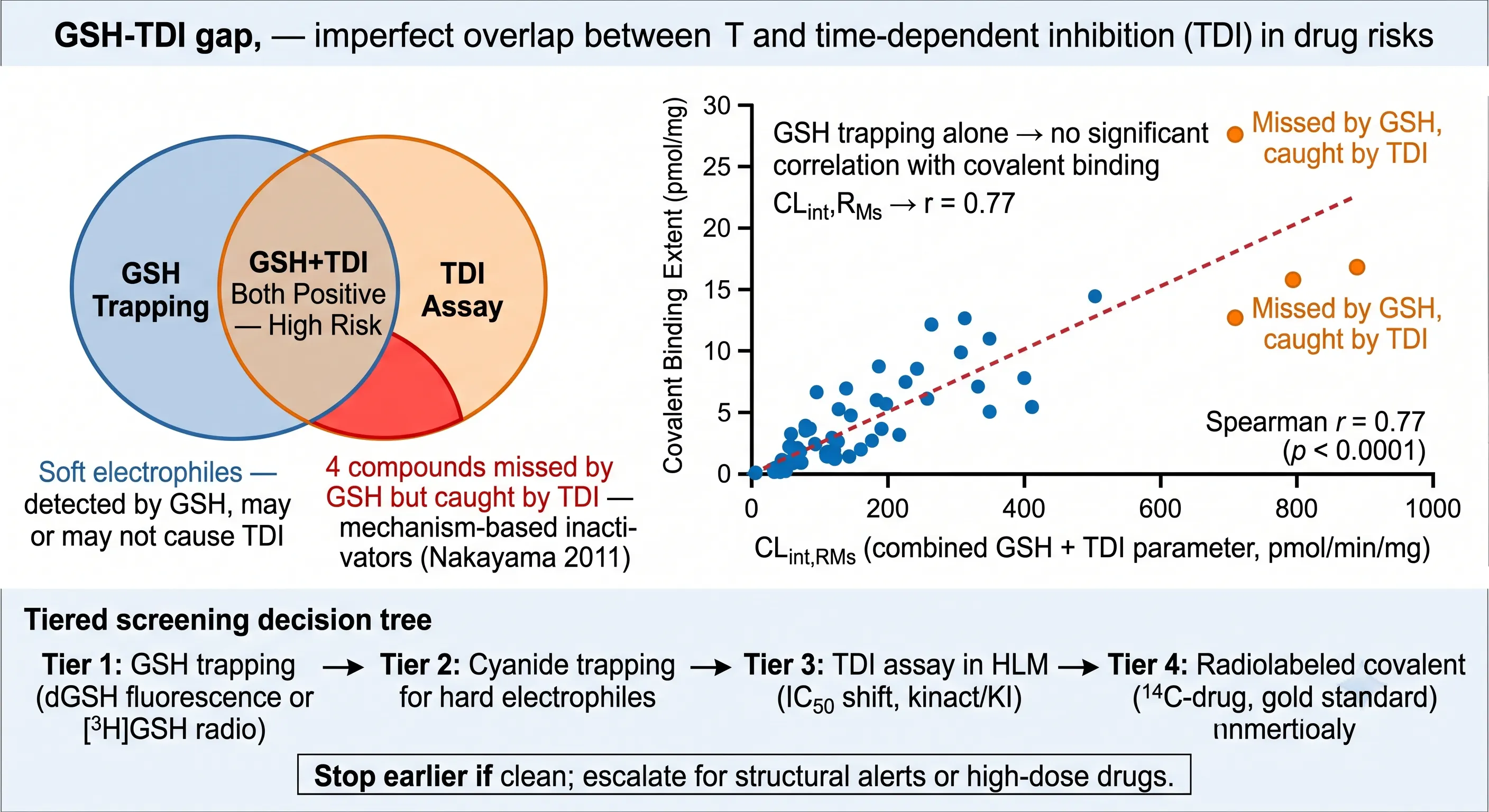 Figure 4: GSH + TDI Combination Strategy — The CLint,RMs Parameter
A reactive metabolite that inactivates the CYP enzyme that formed it is a mechanism-based inhibitor (MBI), and time-dependent CYP inhibition (TDI) is the primary assay for detecting MBIs. The connection between TDI and RM screening is not incidental — the same electrophilic reactive metabolite that forms a GSH adduct can also covalently modify the CYP active site. Integrating GSH trapping and TDI data into a single parameter captures both the "detectable electrophile" (GSH adducts) and the "functional electrophile" (CYP inactivation), providing a more complete picture of RM burden than either assay alone.
Nakayama et al. (2011) formalized this integration as CLint,RMs — a combined intrinsic clearance parameter incorporating both GSH adduct formation and TDI potency — and validated it against the radiometric covalent binding gold standard using [14C]-labeled drugs incubated with HLM (Drug Metab Dispos, 39:392-399). The results were striking: GSH trapping alone showed no statistically significant correlation with covalent binding extent, whereas CLint,RMs achieved a Spearman correlation coefficient of r = 0.77 (p < 0.0001). Four compounds were explicitly identified as "missed by GSH, caught by TDI" — they were mechanism-based inactivators that did not form stable GSH-trappable adducts but nevertheless covalently modified CYP enzymes, producing a positive TDI signal. These four compounds highlight a critical blind spot: a negative GSH trapping result does not rule out reactive metabolite formation if TDI has not also been assessed.
The practical tiered screening strategy that emerges from this data is: (1) GSH trapping (dGSH fluorescence for throughput or [3H]GSH for sensitivity) as the primary screen for soft electrophiles; (2) cyanide trapping for hard electrophiles, particularly for amines and N-heterocycles where iminium ion formation is mechanistically plausible; (3) TDI assay (IC50 shift with 30-minute NADPH pre-incubation, kinact/KI determination) as an orthogonal functional readout that also serves as a DDI risk assessment; and (4) radiometric covalent binding assay using [14C]-labeled drug as the definitive gold standard for compounds that advance to candidate nomination. The decision to escalate is governed by a "stop earlier if clean" principle: if GSH trapping is negative and no structural alerts are present, cyanide trapping and TDI may be deferred; if GSH trapping is positive or structural alerts are present, the full cascade is triggered.
The Amberntsson et al. (2025) study on targeted covalent inhibitors (TCIs) adds an important safety nuance to this framework. Testing six marketed acrylamide-based TCIs plus a proprietary Compound 35 in 3D hepatocyte spheroids, they found that GSH depletion was not the driver of cytotoxicity for any of the seven compounds — ATP depletion preceded toxicity, and none showed GSH-dependent potentiation (Toxicol Res, 14:tfaf054). The practical message: electrophilic warhead reactivity — the designed, target-engagement chemistry of TCIs — should be assessed separately from CYP-mediated oxidative bioactivation. A TCI that is designed to react with a cysteine residue on its target kinase will also react with GSH in a trapping assay, but this is a property of the warhead, not a metabolic liability. The GSH trapping result for a TCI must be interpreted in the context of the warhead's intrinsic reactivity, not taken at face value as an RM risk flag.
Structural Alert Assessment and Risk Stratification Framework
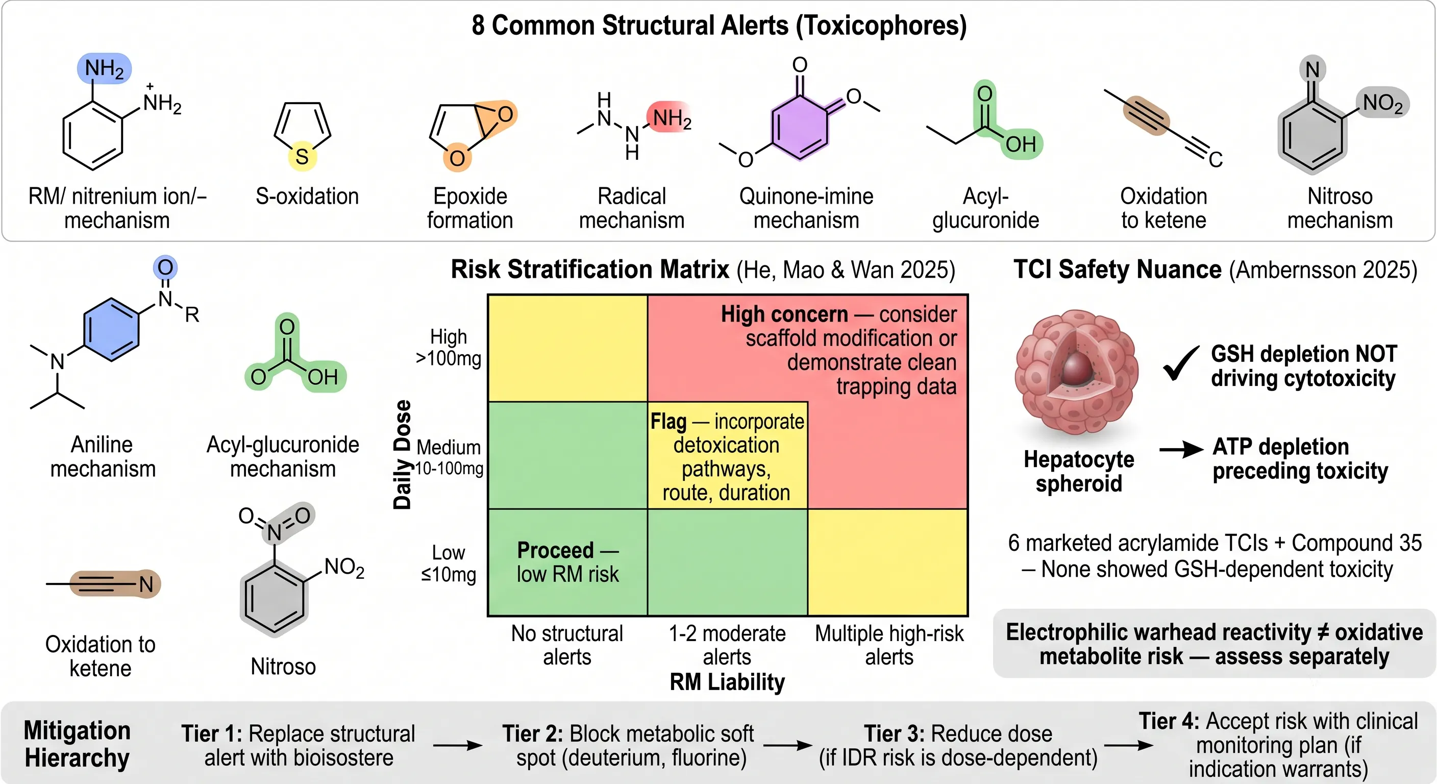 Figure 5: Structural Alert Assessment and Risk Stratification Framework
Before any trapping experiment is run, the compound's structure itself provides actionable RM risk information. Certain functional groups — structural alerts or toxicophores — are mechanistically predisposed to CYP-mediated bioactivation, and their presence in a lead series should trigger proactive RM assessment rather than reactive investigation after toxicity emerges. He, Mao & Wan (2025) comprehensively catalogued the bioactivation mechanisms of organic functional groups, providing the most current reference for structure-based RM risk assessment (Drug Metab Rev, 2025).
The eight highest-priority structural alerts, together with their bioactivation mechanisms, are: aniline (CYP/N-acetyltransferase-mediated N-hydroxylation to nitrenium ion — the classic myeloperoxidase-dependent mechanism implicated in clozapine agranulocytosis); thiophene (CYP-mediated S-oxidation to reactive thiophene-S-oxide or epoxide intermediates); furan (CYP-catalyzed oxidation to furan epoxide, a potent hepatotoxic electrophile); hydrazine/hydrazide (radical-mediated bioactivation producing reactive carbon-centered radicals); p-aminophenol (two-electron oxidation to quinone-imine — the acetaminophen toxicity mechanism at overdose); carboxylic acid (UGT-mediated acyl-glucuronide formation with subsequent acyl migration and protein adduction); terminal alkyne (CYP oxidation to highly reactive ketene); and nitroaromatic (nitroreductase-mediated reduction to nitroso and hydroxylamine intermediates). Each alert carries a known bioactivation mechanism, which means the site of metabolic liability can be targeted for structural modification — the alert is not just a hazard flag, it is a design instruction.
The risk stratification matrix integrates structural alert burden with clinical dose projections. The Y-axis is daily dose (Low ≤10 mg/day; Medium 10-100 mg/day; High >100 mg/day), and the X-axis is RM liability (No structural alerts; 1-2 moderate alerts; Multiple high-risk alerts). The green zone (low dose, clean structure): proceed with standard RM screening — low probability of IDR. The yellow zone (mid-dose, moderate alerts): flag for careful RM assessment; incorporate detoxication pathway analysis, intended route of administration, and treatment duration into the risk calculus. The red zone (high dose, multiple high-risk alerts): high concern — consider scaffold modification (bioisostere replacement) or demonstrate clean double-trapping data with TDI confirmation before advancing. This matrix operationalizes the Uetrecht (2026) dose-dependence principle: a structural alert on a 5 mg/day drug is a manageable risk; the same alert on a 300 mg/day drug is a probable liability.
The mitigation hierarchy for RM-positive compounds follows four tiers: Tier 1 — Replace the structural alert with a bioisostere (e.g., replace aniline with a bioisosteric heterocycle lacking the NH2 group; replace thiophene with a non-sulfur heterocycle of similar shape). Tier 2 — Block the metabolic soft spot (deuterium at the position of CYP oxidation; fluorine or chlorine substitution to sterically or electronically disfavor oxidation). Tier 3 — Reduce the clinical dose if the IDR risk is dose-dependent and the therapeutic window permits it. Tier 4 — Accept the risk with a clinical monitoring plan if the indication warrants it (e.g., oncology, where elevated IDR risk may be acceptable given the disease severity). This hierarchy ensures that RM screening data is actionable — the assay result is not an endpoint but an input to the medicinal chemistry design cycle.
The broader context of drug degradation and instability assessment is covered in forced degradation studies, where chemical degradation pathways are systematically profiled — the same hydrolytic, oxidative, and photolytic mechanisms that produce pharmaceutical degradants can also produce reactive intermediates when they occur enzymatically in vivo, and degradant structural elucidation by HRMS uses MS2 fragmentation logic directly applicable to reactive metabolite adduct characterization.
From Screening Data to Candidate Selection — The Reactive Metabolite Risk Decision Tree
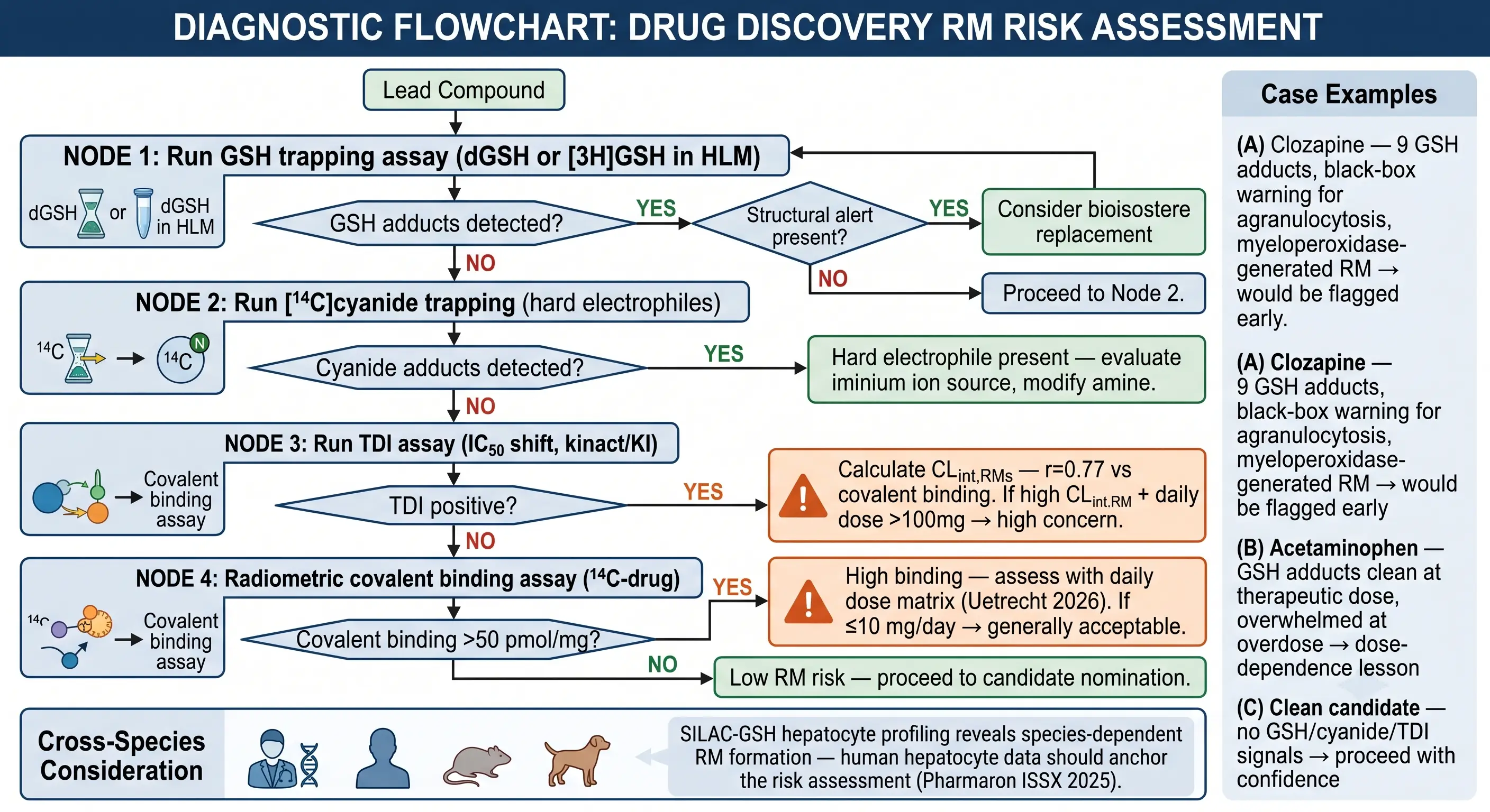 Figure 6: From Screening Data to Candidate Selection — The Reactive Metabolite Risk Decision Tree
The decision tree that translates RM screening data into candidate selection decisions is hierarchical: each node is a gate, and a clean result at any node allows the compound to advance without further RM investigation. The full cascade is reserved for compounds that trigger positive signals at each stage.
Node 1 — GSH trapping (dGSH fluorescence or [3H]GSH radio-HPLC). If no GSH adducts are detected and the compound carries no structural alerts, proceed — RM risk is low. If GSH adducts are detected and a known structural alert explains the bioactivation, consider bioisostere replacement to eliminate the toxicophore before committing resources to downstream RM characterization. If GSH adducts are detected but no structural alert explains the bioactivation, characterize the adduct structurally — the site of GSH conjugation reveals a metabolic soft spot that may not correspond to a classical toxicophore but is equally actionable for medicinal chemistry. If GSH trapping is negative but the compound contains an amine or N-heterocycle, proceed to Node 2 — the negative result may be a false negative for hard electrophiles, not a true negative for RM formation.
Node 2 — Cyanide trapping ([14C]cyanide or CysGlu-Dan fluorescence). If cyanide adducts are detected, the compound forms hard electrophiles (iminium ions or related species). Evaluate the amine substructure generating the electrophile and modify: reduce amine basicity, introduce steric hindrance at the α-carbon position, or replace the secondary/tertiary amine with a metabolically stable bioisostere. If cyanide trapping is negative and Node 1 was also negative, proceed with confidence — both soft and hard electrophile formation are below the detection threshold.
Node 3 — TDI assay (IC50 shift, kinact/KI). A positive TDI result indicates that the reactive metabolite — whether or not it is trapped by GSH or cyanide — is chemically competent to inactivate CYP enzymes. Calculate CLint,RMs (Nakayama 2011 method): if CLint,RMs is elevated and the projected daily dose exceeds 100 mg/day, the compound carries both RM and DDI risk — a double liability that should trigger serious reconsideration. If TDI is negative and both trapping nodes are negative, the RM risk profile is clean.
Node 4 — Radiometric covalent binding ([14C]-drug, gold standard). This is the definitive assay for compounds reaching candidate nomination. If covalent binding exceeds 50 pmol/mg protein, assess against the daily dose matrix (Uetrecht 2026): at ≤10 mg/day, elevated binding is generally acceptable; at 10-100 mg/day, it warrants careful review of the structural alert profile and the TDI data; at >100 mg/day, it constitutes a high-concern finding. If covalent binding is below 50 pmol/mg and all preceding nodes are clean, the RM risk profile supports candidate nomination.
Three real-world cases illustrate the decision tree in practice. Clozapine: 9 GSH-trapped adducts detected via Orbitrap HRMS with FISh scoring, including one unexpected nitrenium ion adduct — the nitrenium ion, generated by myeloperoxidase in neutrophils, is the species responsible for clozapine-induced agranulocytosis. This drug would be flagged at Node 1 in a modern screening cascade and either deprioritized or advanced with a targeted structural modification to block nitrenium ion formation. Acetaminophen: GSH trapping is clean at therapeutic doses (GSH capacity exceeds the quinone-imine formation rate), but overwhelmed at overdose — a demonstration that dose matters as much as intrinsic reactivity. The lesson is not that GSH trapping is unreliable but that RM risk assessment must be dose-contextualized. A structurally clean, low-dose candidate: no GSH adducts, no cyanide adducts, TDI negative — proceed to candidate nomination with confidence. The RM screening cascade exists to identify the clozapines and deprioritize them, not to create barriers for clean compounds.
Cross-Species Considerations and Implementation Strategy
The RM screening data that matters most for regulatory decision-making is the human data — but cross-species comparison provides essential context for toxicology study interpretation. If a reactive metabolite is formed in human hepatocytes but not in rat or dog hepatocytes, the standard two-species toxicology program does not cover the RM-mediated toxicity risk, creating a regulatory gap that is difficult to close at the IND stage.
Pharmaron presented a methodologically elegant solution at ISSX 2025: SILAC-GSH hepatocyte profiling — stable isotope labeling by amino acids in cell culture (SILAC) incorporating labeled glycine into newly synthesized GSH in primary hepatocytes. This enables automated, species-specific detection of GSH adducts without requiring exogenous labeled GSH, providing a direct comparison of RM formation across human, rat, and dog hepatocytes under physiologically relevant GSH homeostasis. The method revealed species-dependent RM formation patterns that would have been missed by standard HLM + exogenous GSH trapping, reinforcing the recommendation that human hepatocyte data — not HLM data — should anchor the RM risk assessment for regulatory submissions.
The operational implementation timeline integrates RM screening into the existing DMPK cascade without adding significant cycle time. At hit-to-lead: in silico structural alert assessment (instant). At lead optimization: dGSH or CysGlu-Dan fluorescence trapping in HLM (1-2 weeks per compound). At late lead optimization: [3H]GSH + [14C]cyanide double trapping with radio-HPLC (2-3 weeks, reserved for compounds with positive primary screens or structural alerts). At candidate nomination: TDI assay + radiometric covalent binding with [14C]-drug (3-4 weeks, one compound). The cumulative data package — structural alert profile, soft electrophile trapping, hard electrophile trapping, TDI assessment, covalent binding quantitation, and cross-species hepatocyte comparison — constitutes a comprehensive RM risk assessment that addresses both the hapten (covalent binding) and danger (cellular stress/TDI) pathways, is interpretable by regulatory reviewers, and most importantly, is actionable by medicinal chemists who can use the site-of-bioactivation information to design safer compounds in the next iteration.
For the same integrated DMPK strategy that connects reactive metabolite risk to the full in vitro ADME profile, advanced drug metabolite identification covers the HRMS workflows for structural characterization of both stable and reactive metabolites, and CYP-mediated drug-drug interaction assessment addresses the TDI component that feeds into CLint,RMs calculation and the risk decision tree at Node 3.
Frequently Asked Questions
What is the difference between the hapten hypothesis and the danger hypothesis of idiosyncratic drug reactions?
The hapten hypothesis (Signal 1) proposes that reactive metabolites covalently bind to cellular proteins, forming drug-protein adducts that are processed and presented as neoantigens on MHC-I, triggering CD8+ T cell-mediated hepatocyte killing — this explains the HLA-dependent, patient-specific nature of IDRs. The danger hypothesis (Signal 2) proposes that reactive metabolites cause cellular stress — mitochondrial damage, ROS release, ER stress, and DAMP release (HMGB1, ATP) — which activates APCs through TLR4/P2X7 receptors, providing the co-stimulatory signal (CD80/CD86) required for full T cell activation. Uetrecht (2026) argues that Signal 2 is NOT idiosyncratic and may be a more reliable predictor of IDR risk than covalent binding alone. Modern RM risk assessment incorporates both pathways: GSH/cyanide trapping assesses covalent binding potential (Signal 1), while TDI and cellular stress assays assess danger signaling potential (Signal 2). The two hypotheses are complementary, not competing — an IDR likely requires both a neoantigen (Signal 1) and a danger signal (Signal 2) to overcome immune tolerance.
Why is single GSH trapping insufficient for comprehensive reactive metabolite detection?
GSH is a soft nucleophile that preferentially traps soft electrophiles — α,β-unsaturated carbonyls, quinones, epoxides, and Michael acceptors. It does NOT efficiently trap hard electrophiles such as iminium ions, which have small, localized positive charges that the diffuse, polarizable thiolate of GSH cannot attack effectively. The 25-drug study by Kawachi et al. (2025) demonstrated that approximately 50% of RM-forming drugs are partially or fully missed by GSH-only trapping: 8 drugs were exclusively GSH-trapped, while 11 required cyanide trapping or both methods for complete detection. The combined GSH + cyanide double trapping, using [3H]GSH for soft electrophiles and [14C]cyanide for hard electrophiles with dual-channel radio-HPLC detection, is now considered the standard for comprehensive electrophile coverage. The RM formation rate × daily dose product (>2,600 pmol/min/mg × mg/day) provided near-complete separation of high-risk from safe drugs in the Kawachi study.
How does the CLint,RMs parameter improve covalent binding risk prediction compared to GSH trapping alone?
CLint,RMs combines GSH trapping data with time-dependent CYP inhibition (TDI) into a single intrinsic clearance parameter. Nakayama et al. (2011) demonstrated that GSH trapping alone showed no significant correlation with covalent binding extent, whereas CLint,RMs achieved a Spearman correlation coefficient of r = 0.77 (p < 0.0001) against the radiometric covalent binding gold standard. Four compounds were explicitly identified as missed by GSH but caught by TDI — these are mechanism-based inactivators that covalently modify CYP enzymes without forming stable, detectable GSH adducts. The practical implication is that TDI screening should be performed alongside GSH trapping as part of an integrated RM assessment, not deferred to a later stage. The combination captures both the electrophiles that form stable GSH adducts (directly detectable) and those that functionally inactivate CYP enzymes (detectable only through TDI), providing a more complete picture of the total RM burden.
What daily dose threshold is associated with low idiosyncratic drug reaction risk?
Drugs administered at ≤10 mg/day rarely cause idiosyncratic drug reactions (IDRs). Uetrecht (2026) systematically reviewed the IDR literature and found that the daily dose is the single most discriminating risk factor: the overwhelming majority of drugs withdrawn or given black-box warnings for IDRs had daily doses exceeding 100 mg. This is incorporated into the He, Mao & Wan (2025) risk stratification matrix as the Y-axis. The mechanistic basis is twofold: lower total body burden of reactive metabolite formation reduces the probability of reaching the threshold for immune activation, and lower dose means fewer drug-protein adducts formed per unit time, making it less likely that any single adduct reaches the antigen presentation threshold. Dose reduction is a valid RM risk mitigation strategy — if a high-RM-liability scaffold can deliver efficacy at ≤10 mg/day, the RM risk is manageable even without structural modification.
What are the most common structural alerts for reactive metabolite formation?
He, Mao & Wan (2025) comprehensively reviewed bioactivation mechanisms of organic functional groups. Eight high-priority structural alerts (toxicophores) are: (1) aniline — CYP/NAT-mediated nitrenium ion formation (implicated in clozapine agranulocytosis); (2) thiophene — CYP-mediated S-oxidation to reactive sulfoxide/epoxide; (3) furan — CYP-catalyzed epoxide formation; (4) hydrazine/hydrazide — radical-mediated bioactivation; (5) p-aminophenol — quinone-imine formation (acetaminophen mechanism at overdose); (6) carboxylic acid — acyl-glucuronide formation with acyl migration and protein adduction; (7) terminal alkyne — CYP oxidation to ketene; (8) nitroaromatic — nitroreductase-mediated reduction to nitroso intermediates. These alerts should be assessed during hit-to-lead using in silico tools, with bioisostere replacement as the first-line mitigation strategy. The mitigation hierarchy is: replace the alert with a bioisostere → block the metabolic soft spot (deuteration, fluorination) → reduce the clinical dose → accept risk with monitoring (oncology or orphan indications only).
At what covalent binding level is a drug candidate considered high-risk?
A radiometric covalent binding level exceeding 50 pmol/mg protein in human liver microsomes is the generally accepted threshold for elevated concern, but this is a contextual decision criterion, not a standalone go/no-go trigger. Uetrecht (2026) emphasizes that multiple safe, widely prescribed drugs exhibit covalent binding well above 50 pmol/mg — covalent binding alone is an imperfect predictor of IDR risk. The interpretation framework integrates covalent binding with daily dose: >50 pmol/mg at ≤10 mg/day is generally acceptable; >50 pmol/mg at 10-100 mg/day warrants careful review of structural alerts and TDI data; >50 pmol/mg at >100 mg/day is a high-concern finding that should trigger scaffold modification or program termination. The GSH + TDI combination parameter (CLint,RMs) provides a more predictive, earlier-stage alternative to radiometric data that correlates with covalent binding (r = 0.77) and can inform compound prioritization before committing to [14C]-drug synthesis.
References
- Uetrecht J. Relevance of reactive metabolites and covalent binding to drug candidate selection. Drug Metab Rev. 2026. DOI: 10.1080/03602532.2026.2620682
- Kawachi T, et al. A novel quantitative assessment of formed reactive metabolites by double trapping with [3H]glutathione and [14C]cyanide. Drug Metab Pharmacokinet. 2025;65:101504. DOI: 10.1016/j.dmpk.2025.101504
- Nakayama S, Atsumi R, Takakusa H, et al. A zone classification system for risk assessment of idiosyncratic drug toxicity using daily dose and covalent binding. Drug Metab Dispos. 2009;37(9):1970-1977. DOI: 10.1124/dmd.109.027797
- Nakayama S, Takakusa H, Watanabe A, et al. Combination of GSH trapping and time-dependent inhibition assays as a surrogate marker of covalent binding. Drug Metab Dispos. 2011;39(3):392-399. DOI: 10.1124/dmd.111.039180
- He Y, Mao Y, Wan H. Bioactivation mechanisms of organic functional groups: structural alerts for reactive metabolite formation. Drug Metab Rev. 2025. DOI: 10.1080/03602532.2025.2472076
- Amberntsson S, Foster AJ, Chouhan B, et al. Use of new approach methodology for hepatic safety assessment of covalent inhibitor drug candidates. Toxicol Res. 2025;14(3):tfaf054. DOI: 10.1093/toxres/tfaf054
- Shibazaki C, Ohe T, Takahashi K, Nakamura S, Mashino T. Development of fluorescent-labeled trapping reagents based on cysteine to detect soft and hard electrophilic reactive metabolites. Drug Metab Pharmacokinet. 2021;39:100386. DOI: 10.1016/j.dmpk.2021.100386
- Pharmaron. SILAC-GSH Hepatocyte Profiling for Species-Dependent Reactive Metabolite Identification. Presented at ISSX 2025. SIL-glycine incorporation into hepatocyte GSH enables automated, species-specific MIP recognition for human/rat/dog comparison.
- ICH M3(R2): Guidance on Nonclinical Safety Studies for the Conduct of Human Clinical Trials and Marketing Authorization for Pharmaceuticals. International Council for Harmonisation; 2009. https://database.ich.org/sites/default/files/M3_R2__Guideline.pdf
- U.S. Food and Drug Administration. Safety Testing of Drug Metabolites: Guidance for Industry (Revision 2). FDA; 2020. https://www.fda.gov/media/72279/download
Related Services


